










Article Contents
Article ID: CM2601113003
Views: 43Method Suitability and Sterility Test Validation of Ciprofloxacin Lactate Solution for Infusion: Overcoming Antimicrobial Activity via Membrane Filtration with Magnesium Sulfate
 PDF
PDF
⬇ Downloads: 1
1Microbiology Laboratory, Profarma Sh.a Pharmaceutical Company, Tirana, Albania
Received: 16 May, 2026
Accepted: 25 September, 2026
Revised: 17 September, 2026
Published: 14 October, 2026
ABSTRACT:
Introduction: Ciprofloxacin inhibits microbial growth and can potentially lead to false-negative sterility results if not adequately neutralized. A validation study was conducted to confirm the suitability of the sterility testing method for ciprofloxacin lactate solution for infusion, with particular emphasis on overcoming the drug’s antimicrobial activity during testing.
Materials and Methods: Magnesium sulfate was evaluated as a chemical inactivator because it forms complexes with fluoroquinolones and reduces their antimicrobial effects. The validation followed harmonized pharmacopeial sterility testing principles (USP ˂71˃, EP 2.6.1), including method suitability trials and representative challenge microorganisms. Test samples containing ciprofloxacin lactate injection were treated with magnesium sulfate before inoculation into Fluid Thioglycolate Medium and Soybean-Casein Digest Medium. Growth promotion, inhibition, and neutralization controls were used to assess the inactivator’s effectiveness.
Results and Discussion: The 1M magnesium sulfate solution and Fluid D successfully neutralized the antimicrobial activity of ciprofloxacin lactate, allowing the recovery and robust growth of all test microorganisms within acceptable timeframes. No microbial growth was observed in the presence of the drug inactivator.
Conclusion: Magnesium sulfate is a reliable and effective inactivator for testing the sterility of ciprofloxacin lactate solution for infusion. The validated method demonstrates accuracy, compliance, and sensitivity to regulatory sterility requirements and supports its routine use for product quality control.
Keywords: Ciprofloxacin lactate, solution for infusion, soybean-casein, sterility testing, magnesium sulfate, antimicrobial activity.
1. INTRODUCTION
Parenteral pharmaceutical products must meet sterility requirements to ensure patient safety, as they bypass the body’s natural defense system [1, 2]. Ciprofloxacin lactate is an injection used to treat systemic infections. It must meet sterility as a quality attribute as such contamination can cause complications, including sepsis and infections in vulnerable populations [3].
Sterility testing is an important quality control step for injectable formulations. Pharmacopoeial standards, such as the United States Pharmacopeia and European Pharmacopeia (European Directorate for the Quality of Medicines), recommend two main approaches: direct inoculation and membrane filtration [2, 4]. Membrane filtration is preferred for aqueous solutions and products with antimicrobial properties, as it removes inhibitory substances by concentrating specific microbial contaminants on a filter that can be incubated in suitable growth media [5].
Residual antibacterial activity in the ciprofloxacin formulation can affect sterility test results by inhibiting microbial growth, resulting in a false-negative outcome [6, 7]. This supports validation of the sterility testing method, including suitability for effective neutralization or removal of any antimicrobial agent and the ability to recover low levels of microorganisms, as per the EDQM and USP [2, 4].
This study focused on validating the membrane filtration method for sterility testing of ciprofloxacin lactate solution for infusion. Validation helps evaluate key parameters such as microbial recovery, method suitability, and the effectiveness of the rinsing procedure in eliminating antibacterial activity, based on pharmacopeia guidelines and international standards such as the International Council for Harmonization [2, 8]. This study investigated whether adding the neutralizing agent Mg2+ during sterility testing can eliminate the test sample’s antibacterial activity, as Mg2+ forms complexes with quinolone drugs. Establishing a reliable and validated sterility testing method is essential to ensure compliance with regulatory standards and guarantee the microbiological safety of the final product [9].
2. MATERIALS AND METHODS
2.1. Reagents and Instrumentations
In this study, the following reagents and instruments were used: Analytical balance Explorer OHAUS, Euroclone Top safe microbiology safety cabinet, Petri plates Isolab, IKA MS 3 Basic Vortex, Tuttnauer model 3850 MLV, INE 800 Memmert incubator, UM 800 Memmert incubator, pH meter Ultrabasic UB-10, Denver Instrument, Millipore Steritest Compact Pump (Merck), Steritest Devices for antibiotics (Merck Millipore).
Three batches of ciprofloxacin solution for infusion Cat no. 2404; 2502; 2505 was manufactured in Profarma Sh.a), Trypticasein Soy Agar EP/USP/ISO (medium Cat No.1068 by Condalab, Spain), Sabouraud Dextrose Agar 4% medium (Cat. No.1024 by Condalab, Spain); Fluid A (Cat. No. 638042) and Fluid D (Cat. no. 638043), Thioglycolate Medium (FTM, Cat. No. 638040 and Soybean-Casein Digest Medium (SBCDM, Cat. No. 622787) were purchased from Merck Millipore, USA); Lyophilized bacterial cultures (containing 10-100 CFU/ml) of Staphylococcus aureus (ATCC 6538), Bacillus subtilis (ATCC 6633), Pseudomonas aeruginosa (ATCC 9027), Clostridium sporogenes (ATCC 19404), and fungal strains Candida albicans (ATCC 10231) and Aspergillus brasiliensis (ATCC 16404) were obtained as EZ- Accu Shot ™ quantitative microorganism preparation (Microbiologics, Inc., St. Cloud, MN, USA) and were prepared according to the manufacturer’s instructions to obtain the challenge inoculum. The microorganism Micrococcus luteus was an environmental isolate collected from the cleanrooms to organize the tests.
2.2. Sterilization
All sterility tests were performed aseptically in ISO Class 5 Biological Safety Cabinet (BSC), providing a controlled environment to minimize the risk of environmental contamination during sample handling and filtration [2, 4, 10, 11]. Sterilization of Trypticasein Soy Agar and Sabouraud Dextrose Agar 4% was performed at 121˚C for 15 minutes using a steam autoclave. Sterility testing of ciprofloxacin lactate solution for infusion was performed using the membrane filtration method in accordance with the requirements of the United States Pharmacopeia and the European Pharmacopeia (European Directorate for the Quality of Medicines and HealthCare [2, 4]. The procedure was carried out under aseptic conditions using sterile membrane filters with a nominal pore size of 0.45 micrometer. A defined volume of sample was filtered, followed by thorough rinsing of the membrane with sterile diluent to remove any residual antimicrobial activity. The membrane was transferred into specific culture media, that is, fluid thioglycolate medium and soybean casein medium, incubated at a specific temperature for at least 14 days, and the media were examined for microbial growth. Appropriate positive controls, negative controls, and growth promotion controls were included to confirm the validity of the test and the suitability of the procedure. The suitability, sterility, and sensitivity tests of the culture media were conducted in accordance with Ph. Eur. 2.6.1.
2.3. Growth Media Control
Sterility testing was performed using compendial culture media suitable for detecting a broad range of microorganisms. Before validation of the sterility test method, growth medium control was used to verify the suitability of the culture media. Tryptic Soy Agar (TSA) and Sabouraud Dextrose Agar 4% (SDA) are solid media used for growth promotion, colony isolation, and confirming the viability of challenge organisms used in validation. They were examined for sterility and growth-promoting properties. Each batch of medium was visually inspected for its appearance, pH, color, and incubated under appropriate conditions to confirm sterility.
Growth promotion testing was carried out using appropriate reference microorganisms to demonstrate TSA’s ability to support aerobic bacterial growth for up to 3 days at 35 °C and yeast and mold growth for up to 5 days at 25 °C. Fluid Thioglycolate Medium (FTM) was used for the cultivation of anaerobic and aerobic bacteria. It was incubated at 30 to 35 °C, whereas Soybean-Casein Digest Medium (SCDM) was used to detect aerobic bacteria and fungi and was incubated at 20 to 25 °C. Both media were purchased from Merck, an approved supplier. They underwent growth promotion testing before using to confirm their ability to support the growth of specified indicator microorganisms. Media sterility controls were included to verify the absence of contamination before testing and ensure the validity and reliability of the sterility test results.
Tables 1 and 2 represent the detail of media and microbial cultures used in the validation method. These procedures included the measurement of pH checks to ensure that the media pH was within specifications. We assigned a batch number and shelf life to all media and maintained batch manufacturing documentation.
Table 1. Recommended microbial cultures for TSA and SDA.
| Name of the Media | Microbial Cultures Recommended | Incubation Temperature (0C) | |
| Aerobic Bacteria: | |||
| Tryptic Soy Agar (TSA) | Staphylococcus aureus | ATCC 6538 | 30-35 |
| Bacillus subtilis | ATCC 6633 | ||
| Pseudomonas aeruginosa | ATCC 9027 | ||
| Fungi: | |||
| Tryptic Soy Agar (TSA) | Candida albicans | ATCC 10231 | 30-35 |
| Aspergillus brasiliensis | ATCC 16404 | ||
| Fungi: | |||
| Sabouraud Dextrose Agar (SDA) | Candida albicans | ATCC 10231 | 20-25 |
| Aspergillus brasiliensis | ATCC 16404 | ||
Table 2. Media and recommended microbial cultures for sterility testing.
| Name of the Media | Microbial Cultures Recommended | Incubation Temperature (0C) | |
Fluid Thioglycolate Medium (FTG medium/FTM) | Aerobic Bacteria: | 30-35 | |
| Staphylococcus aureus | ATCC 6538 | ||
| Bacillus subtilis | ATCC 6633 | ||
| Pseudomonas aeruginosa | ATCC 9027 | ||
| Anaerobic Bacteria: | |||
| Clostridium sporogenes | ATCC 19404 | ||
| Soybean-Casein Digest Medium (SCDM) | Aerobic Bacteria: | 20-25 | |
| Bacillus subtilis | ATCC 6633 | ||
| Fungi: | 20-25 | ||
| Candida albicans | ATCC 10231 | ||
| Aspergillus brasiliensis | ATCC 16404 | ||
| Soybean-Casein Digest Medium (SCDM) | Environment Isolate | 20-25 | |
| Micrococcus luteus | |||
| Fluid Thioglycolate Medium (FTG medium /FTM) | Environment Isolate | 30-35 | |
| Micrococcus luteus | |||
2.4. Tested Samples
Sixty ampoules from three different batches (20 samples per batch) contained ciprofloxacin lactate solution for infusion (100 mg/10mL) and were selected to ensure representativeness in accordance with pharmacopeial requirements [2, 4]. Before testing, a QC analyst visually inspected the ciprofloxacin lactate ampoules to confirm container-closure integrity and the absence of visible particulate matter. Before testing, the ampoules were inspected for integrity and the absence of visible particulates. The ampoules were properly disinfected, and all manipulations, including opening and transferring the contents, were performed in a laminar airflow cabinet using sterile equipment and aseptic techniques. These precautions helped ensure reliable sterility test results.
2.5. Method Suitability
Method suitability testing was performed to demonstrate that ciprofloxacin lactate (i.e., the infusion solution) had no bacteriostatic or fungistatic effects on the sterility test. The product was inoculated separately with low levels (10-100 CFU/ml) of particular compound-sensitive microorganisms, including anaerobic bacteria, aerobic bacteria, and fungi, and subjected to sterility test procedures. Membrane filtration is followed by validated rinsing steps to minimize antimicrobial interference. The validated rinsing steps included pre-wetting with 50 mL of Fluid A, filtration of 100 mL of ciprofloxacin lactate, and addition of 200 mL of 50 mL of 1 M magnesium sulfate. The final step included rinsing with 200 mL of Fluid D and 100 mL of Fluid A. Thus, a total volume of 500mL was used for each membrane. A total of 20 ampules from each batch were selected for sterility testing, and half of each 5 mL ampule was filtered through each membrane. We compared the growth and recovery of the challenging organism in the presence of the product with that of the positive controls. We used a polyvinylidene difluoride (PVDF) 0.45 µm low-binding filter to effectively trap microorganisms without allowing ciprofloxacin to bind to the filter membrane, which might hinder microorganism recovery. The membrane was pre-wetted with Fluid A before the experiment to condition the membrane, ensure even fluid flow, and reduce product binding. The membrane was then rinsed with Fluid D and Fluid A to eliminate any remaining drugs and inactivating agents.
2.6. Validation Parameters
The sterility testing method was validated using a series of critical parameters to ensure its reliability and regulatory compliance [12–14]. Method suitability testing involved inoculating the test sample with known compendial microorganisms representing aerobic bacteria, anaerobic bacteria, and fungi to confirm the method’s ability to detect low levels of contamination in the presence of the product. We demonstrated neutralization of antibiotic activity by incorporating validated inactivation and rinsing steps and comparing microbial recovery in the presence of the product with that in positive control samples, confirming the absence of bacteriostatic or fungistatic effects [15–17]. Membrane integrity and sterility controls were included to verify the physical integrity of the filtration membrane and ensure that the filtration system, media, and diluents remained sterile throughout the test procedure. In addition, growth promotion testing of the culture media was conducted before use to confirm that the media supported the growth of the specified indicator microorganisms, ensuring the media’s suitability for sterility testing. This traditional approach aligned with the qualitative nature of compendial sterility testing and its validation, where successful recovery of the specified challenge microorganisms was the primary acceptance criterion.
3. RESULTS
3.1. Growth Promotion Testing of TSA and SDA
Growth promotion testing confirmed that Tryptic Soy Agar (TSA) and Sabouraud Dextrose Agar (SDA) supported growth of the specified challenge microorganisms. Each microorganism was inoculated at a concentration of 10-100 cfu/ml. All test microorganisms showed visible growth after incubation, with colony morphology consistent with each organism’s expected characteristics (Tables 3 and 4). No microbial growth was detected in the uninoculated control media (negative control, NC), confirming the sterility of the prepared media. These findings show that both TSA and SDA met the acceptance criteria for growth promotion testing and confirm the viability of the challenge organisms used in the validation.
Table 3. Observations of microbial growth of challenged microorganisms on TSA at 35°C.
| Culture/Media | Description | Day of Observation | ||
| Day 1 | Day 2 | Day 3 | ||
| S. aureus/TSA | PC | – | + | + |
| P. aeruginosa/TSA | PC | – | + | + |
| B. subtilis/TSA | PC | + | + | + |
| Candida albicans/TSA | PC | – | – | + |
| Aspergillus brasiliensis /TSA | PC | – | + | + |
| Negative control | NC | – | – | – |
Keys: PC-Positive Control; +: Growth observed; -: Growth not observed
Table 4. Observations of microbial growth of challenged microorganisms on SDA at 25°C.
| Culture/Media | Description | Date of Observation | ||||
| Day 1 | Day 2 | Day 3 | Day 4 | Day 5 | ||
| Candida albicans/SDA | PC | – | – | + | + | + |
| Aspergillus brasiliensis/SDA | PC | – | – | + | + | + |
| Negative control | NC | – | – | – | – | – |
Keys: PC-Positive Control; +: Growth observed; -: Growth not observed
3.2. Results of Bacteriostasis and Fungistasis Studies
Bacteriostasis and fungistasis Testings demonstrated the successful recovery of all specified bacterial and fungal test organisms in the presence of the product following membrane filtration and appropriate neutralization. Visible bacterial growth was observed within the prescribed incubation period in Fluid Thioglycolate Medium (Table 5), whereas characteristic fungal growth was detected in Soybean-Casein Digest Medium (Table 6). No inhibition of microbial growth was observed compared to the positive controls, and no growth was detected in the negative controls. These results confirm the absence of bacteriostatic and fungistatic effects and validate the sterility testing method for the reliable detection of microbial contamination.
Table 5. Bacteriostasis study results.
| Culture | Description | Day of Observation | ||
| 1st | 2nd | 3rd | ||
| S. aureus | PC | – | + | + |
| PPC | – | + | + | |
| B. subtilis | PC | – | + | + |
| PPC | – | + | + | |
| P. aeruginosa | PC | – | + | + |
| PPC | – | + | + | |
| C. sporogenes | PC | – | + | + |
| PPC | – | + | + | |
| Micrococcus luteus | PC | – | + | + |
| PPC | – | + | + | |
| Negative control | NC | – | + | + |
Keys: PC – Positive Control; PPC – Positive Product Control
+: Growth observed; -: Growth not observed; NC-negative control
Table 6. Fungistasis study results.
| Culture | Description | Day of Observation | ||||
| 1st | 2nd | 3rd | 4th | 5th | ||
| C. albicans | PC | – | – | + | + | + |
| PPC | – | – | + | + | + | |
| A. brasiliensis | PC | – | + | + | + | + |
| PPC | – | – | + | + | + | |
| Negative control | NC | – | – | – | – | – |
Keys: PC – Positive Control; PPC – Positive Product Control
+: Growth observed; -: Growth not observed; NC- negative control
Visible microbial growth was observed in all inoculated test samples (PPC) and positive controls (PC) on the second day of bacterial incubation. Positive Product Control (PPC) showed identical visual turbidity (Fig. 1) to the Positive Control (PC) within 5 days, while no growth was detected in negative controls, demonstrating the absence of inhibitory effects and confirming the suitability of the membrane filtration method for routine sterility testing of ciprofloxacin lactate solution for infusion.
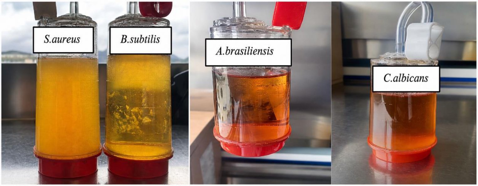
Fig. (1). Growth of bacteria, mold, and fungi on Positive Product Control (PPC) after 3 and 5 days.
The results demonstrated that 1 M magnesium sulfate and Fluid D successfully neutralized the antimicrobial activity of ciprofloxacin lactate and cleared all antimicrobial residues from the membrane, allowing recovery and robust growth of all test organisms within acceptable timeframes. No inhibition of microbial growth was observed in the presence of the drug-inactivator mixture. Negative controls of each medium and test specimens from three batches were examined after 14 days and showed no microbial growth (Table 7). These results confirmed that the three batches of ciprofloxacin lactate solution for infusion were sterile after 14 days of incubation.
Table 7. Sterility test report.
| Day | FTM (Incubation at 30-35ºC) | SCDM (Incubation at 20-25ºC) | ||||||
| Positive Control | Negative Control | Positive Product Control | Test Specimen | Positive Control | Negative Control | Positive Product Control | Test Specimen | |
| 1 | + | – | – | – | – | – | – | – |
| 2 | + | – | + | – | + | – | – | – |
| 3 | + | – | + | – | + | – | + | – |
| 4 | NA | – | NA | – | + | – | + | – |
| 5 | NA | – | NA | – | + | – | + | – |
| 6 | NA | – | NA | – | NA | – | NA | – |
| 7 | NA | – | NA | – | NA | – | NA | – |
| 8 | NA | – | NA | – | NA | – | NA | – |
| 9 | NA | – | NA | – | NA | – | NA | – |
| 10 | NA | – | NA | – | NA | – | NA | – |
| 11 | NA | – | NA | – | NA | – | NA | – |
| 12 | NA | – | NA | – | NA | – | NA | – |
| 13 | NA | – | NA | – | NA | – | NA | – |
| 14 | NA | – | NA | – | NA | – | NA | – |
Note: + = Growth observed; − = Growth not observed; NA = Not applicable after confirmation of adequate microbial growth in the control cultures. After a visual examination for good growth of each microorganism (day 3 for bacteria and day 5th for fungi/mold), canisters were removed from incubators. Negative controls of each medium and test specimen for three lots were examined after 14 days.
The study confirmed that magnesium sulfate is an effective and reliable inactivator for the sterility testing of ciprofloxacin lactate solution for infusion. The validated method ensures accuracy, sensitivity, and compliance with regulatory sterility requirements, supporting its routine use for product quality control.
4. DISCUSSION
Regulatory requirements emphasize that sterility tests must be validated to demonstrate their suitability, particularly for antimicrobial products such as ciprofloxacin. This includes assessing bacteriostatic and fungistatic effects to ensure the method does not inhibit microbial growth, which could lead to false-negative results [2, 4]. Method suitability testing must confirm the recovery of viable microorganisms in the presence of the product, demonstrating effective neutralization or removal of the antimicrobial activity [6, 7]. Validation must include growth promotion testing, appropriate positive and negative controls, and specified challenge microorganisms, with incubation for at least 14 days. GMP and ICH guidelines require sterility testing and method suitability validation to ensure reliable detection of microbial contamination in sterile products [8, 10, 18, 19].
The sterility test was considered acceptable if all validation parameters met the following criteria: growth of all inoculated microorganisms must be clearly visible in method suitability tests, demonstrating the method’s ability to detect contamination; no significant inhibition of microbial growth should occur compared to positive control samples, confirming effective neutralization of ciprofloxacin’s antimicrobial activity; no growth should be observed in negative controls and test controls, ensuring that contamination did not arise from the test system or materials; and all culture media must support expected growth during growth promotion testing, verifying their suitability for detecting microorganisms. Meeting these acceptance criteria confirmed that the sterility test method was reliable, robust, and suitable for routine testing of ciprofloxacin lactate solution for infusion.
Product formulation characteristics directly influence the sterility testing strategy and method suitability. The preferred sterility test method was membrane filtration using a closed system for several reasons. First, it is generally compatible with membrane filtration because the product is aqueous in nature. Second, membrane filtration allows for easier filtration and thorough rinsing of the membrane. Third, it often supports the more efficient removal of inhibitory substances that can interfere with microbial growth. Therefore, membrane filtration was considered more sensitive and appropriate than direct inoculation.
The antimicrobial nature of ciprofloxacin shows a significant challenge in sterility testing because it can inhibit microbial growth and produce false-negative results. Therefore, we established method suitability in accordance with USP <71> and USP <1227>. We demonstrated suitability by successfully recovering the prescribed challenge microorganisms, confirming both effective neutralization of antimicrobial activity and the neutralizer’s lack of toxicity. USP <1227> recognizes that antimicrobial activity may be neutralized by chemical neutralization, membrane filtration, rinsing, or combinations thereof, provided that the selected approach is validated through method suitability studies. A study by Liu et al. [20] demonstrated that a quinolone injection required optimized membrane filtration, rinsing, and neutralization to eliminate antimicrobial activity during sterility testing [20]. Proper rinsing and filtration techniques significantly improve the detection of viable microorganisms in injectable products [21].
The standard mitigation steps included two neutralizing agents, magnesium sulfate and Fluid D. Initial method suitability testing using 400 mL of Fluid D alone could not neutralize the drug and caused false-negative results. The combination of 0.5 M magnesium sulfate and a rinse with Fluid A failed to eliminate the product’s inherent antimicrobial activity, resulting in persistent fungistasis. By increasing the neutralizing agent concentration to 1 M magnesium sulfate, divalent magnesium (Mg²⁺) ions successfully chelated the residual antibiotic bound to the membrane filter. In this study, the sterility testing of ciprofloxacin lactate (solution for infusion) employed a red-based canister with a PVDF 0.45 µm low-binding membrane, pre-wetting with Fluid A, inactivation with 1 M magnesium sulfate (200 mL), then rinsing with Fluid D (200 mL) and Fluid A (100 mL) with a total rinse volume of 500 mL.
This optimization allowed for robust visual growth of the challenged microorganisms within the mandated 5-day incubation period without exerting osmotic toxicity. Consequently, this modified rinsing protocol neutralizes the formulation’s inhibitory effects and establishes a validated, compliant membrane filtration sterility test under the USP <71> guidelines. Bacteriostasis and fungistasis studies were performed as part of the sterility test method suitability to demonstrate that ciprofloxacin lactate does not inhibit microbial growth, thereby ensuring the reliability of the sterility testing results. In a bacteriostasis study, representative bacterial strains were inoculated at low levels (≤100 CFU) into the test system containing the product, followed by membrane filtration and appropriate rinsing or neutralization. The test was considered acceptable when visible bacterial growth was observed within the prescribed incubation period and was comparable to the growth observed in the positive controls. The presence of growth indicates that the product does not exert bacteriostatic or bactericidal effects that could mask microbial contamination during routine sterility testing. Similarly, a fungistasis study evaluated the product’s potential inhibitory effect on fungi, including yeasts and molds. After exposing the membrane or test medium to the product, the product was inoculated with fungal test organisms and incubated under suitable conditions. Acceptance is demonstrated by the appearance of characteristic fungal growth within the specified timeframe, confirming that the product does not inhibit fungal recovery. The combined results of bacteriostasis and fungistasis studies support the validity of sterility testing by showing that any contaminating bacteria or fungi, if present, would be capable of growth and detection, and the absence of inhibition confirmed the suitability of this method. Method suitability testing with compendial microorganisms demonstrated effective antibiotic neutralization, recovery of all challenged organisms, and compliance with sterility and growth promotion controls. This study confirmed that the combination of magnesium sulfate neutralization and validated rinsing with Fluid A and Fluid D ensures reliable sterility testing of ciprofloxacin lactate solution for infusion by effectively eliminating bacteriostatic and fungistatic interference. Traditional sterility testing methods, including membrane filtration, remain relevant because they are pharmacopeia-approved, dependable, and inexpensive. However, current technologies for sterility testing are more rapid and automated, thus providing prompt results, and require the use of sophisticated technology and validation. Membrane filtration remains a sensible and relevant choice.
CONCLUSION
This study demonstrates that sterility testing of ciprofloxacin lactate solution for infusion can be reliably performed using a PVDF 0.45 µm low-binding membrane with pre-wetting, inactivation with 200 mL of 1 M magnesium sulfate, and a rinse volume of 200 mL of Fluid D and 100 mL Fluid A. Method suitability testing confirmed effective neutralization of ciprofloxacin’s antimicrobial activity, with recovery of all compendial microorganisms and no inhibition observed compared to positive controls. Membrane integrity, sterility controls, and growth promotion tests further validated the procedure’s robustness. Compared with previous studies that used larger rinse volumes, optimizing neutralizer concentration and rinsing medium enabled complete microbial recovery at a reduced solution volume, improving efficiency while preserving testing accuracy. Overall, the technique is a reliable and validated method for assessing the sterility of antibiotic-containing parenteral products.
RECOMMENDATIONS
Sterility testing of antibiotic-containing injections should use neutralization and rinsing procedures to overcome antimicrobial properties, test method suitability with the selected microorganisms, and regularly test membrane integrity and growth promotion. The neutralizer concentration and rinse volumes should be optimized for maximal efficiency.
LIST OF ABBREVIATIONS
FTM | = | Fluid Thioglycolate Medium |
PVDF | = | Polyvinylidene Difluoride |
SDA | = | Sabouraud Dextrose Agar |
SCDM | = | Soybean-Casein Digest Medium |
TSA | = | Tryptic Soy Agar |
AUTHOR’S CONTRIBUTION
E.T. has contributed to the study conceptualization, methodology, data analysis, interpretation of results, and manuscript writing.
ETHICAL APPROVAL AND INFORMED CONSENT
This manuscript does not report on any studies involving animal subjects. All experimental procedures were strictly in vitro laboratory assays conducted using compendial microbiological reference strains in accordance with institutional laboratory guidelines. This report does not include any experiments conducted on live animal subjects; therefore, traditional animal welfare committee approvals are not applicable. All experimental procedures were strictly in vitro laboratory assays executed in complete compliance with the United States Pharmacopeia Method Suitability Guidelines (USP <71> Sterility Testing) and the European Pharmacopoeia (EP 2.6.1). Biological materials were limited to compendial microbiological reference strains managed according to the institutional biosafety and standard operating procedures of the Department of Quality Control Laboratory in Profarma, located in Albania for the safe handling of non-pathogenic microorganisms.
AVAILABILITY OF DATA AND MATERIAL
The data will be made available on reasonable request by contacting the corresponding author [E.T.].
FUNDING
None.
CONFLICT OF INTEREST
The author is a member of the Editorial Board of this journal. The author declares no other conflicts of interest. The manuscript was handled independently, and the author was not involved in the peer-review or editorial decision-making process.
ACKNOWLEDGEMENTS
Declared none.
DECLARATION OF AI
During the preparation of this manuscript, the author used ChatGPT for language editing and refinement purposes. Following the use of this tool, the author carefully reviewed and revised the content where necessary and accept full responsibility for the final published version of the article.
REFERENCES
[1] Adejare A, editor. Remington: The science and practice of pharmacy. 23rd ed. London: Academic Press; 2021.
https://doi.org/10.1016/C2018-0-04991-9
[2] United States Pharmacopeia. General Chapter <71> Sterility tests. In: USP-NF. Rockville (MD): United States Pharmacopeial Convention; 2025.
https://doi.org/10.31003/USPNF_M98810_01_01
[3] Buckingham R, editor. Martindale: The complete drug reference. 40th ed. London: Pharmaceutical Press; 2020. Available from: https://www.ncbi.nlm.nih.gov/nlmcatalog/101779009 (Accessed on: 19-09-2026).
[4] European Directorate for the Quality of Medicines and HealthCare. European Pharmacopoeia. 5th ed. Strasbourg: Council of Europe; 2005. Chapter 2.6.1, Sterility; p. 145–8. Available from: https://www.atlasbio.com/wp-content/uploads/2016/05/EP-2.6.1-Sterility.pdf (Accessed on 19-09-2026).
[5] Denyer SP, Hodges NA, Gorman SP, Gilmore BF, editors. Hugo and Russell’s pharmaceutical microbiology. 8th ed. Chichester: Wiley-Blackwell; 2011. Available from: https://pure.qub.ac.uk/en/publications/hugo-amp-russells-pharmaceutical-microbiology-8th-edition/ (Accessed on: 19-09-2026).
[6] Sandle T. Sterility testing of pharmaceutical products. Bethesda (MD): PDA/DHI; 2013. Available from: https://openlibrary.org/books/OL25432834M/Sterility_Testing_of_Pharmaceutical_Products (Accessed on: 19-09-2026).
[7] Sutton SVW. The most probable number method and its uses in enumeration, qualification, and validation. J Valid Technol. 2010;16(3):35–38. Available from: https://www.americanpharmaceuticalreview.com/Featured-Articles/36986-Activities-of-the-USP-Microbiology-and-Sterility-Assurance-Expert-Committee-During-the-2005-2010-Revision-Cycle/ (Accessed on: 19-09-2026).
[8] International Council for Harmonisation. ICH Q7: Good manufacturing practice guide for active pharmaceutical ingredients. Geneva: International Council for Harmonisation; 2000. Available from: https://database.ich.org/sites/default/files/Q7%20Guideline.pdf (Accessed on: 19-09-2026).
[9] Lehto P, Kivistö KT, Neuvonen PJ. The effect of ferrous sulphate on the absorption of norfloxacin, ciprofloxacin and ofloxacin. Br J Clin Pharmacol. 1994;37(1):82–85.
https://doi.org/10.1111/j.1365-2125.1994.tb04245.x
[10] European Commission. EudraLex, Volume 4: EU guidelines for good manufacturing practice for medicinal products for human and veterinary use, Annex 1: Manufacture of sterile medicinal products. Brussels: European Commission; 2022. Available from: https://health.ec.europa.eu/document/download/e05af55b-38e9-42bf-8495-194bbf0b9262_en (Accessed on 19-09-2026).
[11] Sandle T. Practical approaches to sterility testing. In: Saghee MR, Sandle T, Tidswell EC, editors. Microbiology and sterility assurance in pharmaceuticals and medical devices. New Delhi: Business Horizons; 2011. p. 173–192. Available from: https://openlibrary.org/books/OL24821357M/Microbiology_and_Sterility_Assurance_in_Pharmaceuticals_and_Medical_Devices (Accessed on: 19-09-2026).
[12] Sandle T. Practical approaches to sterility testing. J Valid Technol. 2004;10(2):131–141. Available from: https://www.researchgate.net/publication/298418865_Practical_Approach_To_Sterility_Testing (Accessed on: 19-09-2026).
[13] Sandle T. In 1975, the Steritest™ system made the sterility test reliable: the story of its invention. Pharmaceutical Microbiology Resources; 2020. Available from: https://www.pharmamicroresources.com/2020/11/in-1975-steritest-system-made-sterility.html (Accessed on: 19-09-2026).
[14] Russell AD, Ahonkhai I, Rogers DT. A review: microbiological applications of the inactivation of antibiotics and other antimicrobial agents. J Appl Bacteriol. 1979;46(2):207–245.
https://doi.org/10.1111/j.1365-2672.1979.tb00818.x
[15] Merck Millipore. Sterility testing of products with antimicrobial properties. Darmstadt: Merck Millipore; [date unknown]. Available from: https://www.merckmillipore.com/IL/en/technical-documents/technical-article/microbiological-testing/sterility-testing/sterility-testing-with-antimicrobial (Accessed on: 19-09-2026).
[16] Guillard N, Delaunay C, Olivier E, Cormier N. Development and validation of a sterility test method by membrane filtration of ceftazidime 5% eye drops. Pau: GERPAC; 2021. Available from: https://www.gerpac.eu/development-and-validation-of-sterility-test-method-by-membrane-filtration-of-ceftazidime-5-eye-drops (Accessed on: 19-09-2026).
[17] Akers MJ, Wright GE, Carlson KA. Sterility testing of antimicrobial-containing injectable solutions prepared in the pharmacy. Am J Hosp Pharm. 1991;48(11):2414–2418.
https://doi.org/10.1093/ajhp/48.11.2414
[18] US Food and Drug Administration. Title 21, Code of Federal Regulations, §3.2 (e)(1–2): Definition of combination product. Silver Spring (MD): US Department of Health and Human Services; 2023. Available from: https://www.ecfr.gov/current/title-21/chapter-I/subchapter-A/part-3/section-3.2 (Accessed on: 19-09-2026).
[19] World Health Organization. WHO good manufacturing practices for sterile pharmaceutical products. Geneva: World Health Organization; 2011. (WHO Technical Report Series, No. 961, Annex 6). Available from: https://www.who.int/docs/default-source/medicines/norms-and-standards/guidelines/production/trs961-annex6-gmp-sterile-pharmaceutical-products.pdf (Accessed on: 19-09-2026).
[20] Liu D, Tang C, Hao Z. Establishment and validation of sterility test of levofloxacin lactate and sodium chloride injection. J Pharm Pract Serv. 2015;33(6):552–4.
https://doi.org/10.3969/j.issn.1006-0111.2015.06.019
[21] Pan JW, Guo XQ. Establishment and validation of the sterility test method for ferric pyrophosphate citrate injection. Chin J Pharm Biotechnol. 2017;24(2):141–4.
https://doi.org/10.19526/j.cnki.1005-8915.20170211
Licensed
© 2026 Copyright by the Authors.
Licensed as an open access article using a CC BY 4.0 license.
Article Contents Author Eva Troja1, * 1Microbiology Laboratory, Profarma Sh.a Pharmaceutical Company, Tirana, Albania Article History: Received: 16 May, 2026
Article Contents Author Felicia Gabriela Beresescu1, * 1George Emil Palade University of Medicine, Pharmacy, Science and Technology, Targu Mures, Romania
Article Contents Authors Saman Ismail1, * Imran Iqbal2 1Faculty of Management Science, Department of Business Administration, Salim Habib University, Karachi,
Article Contents Author Michael Dolinsky1, * 1Faculty of Mathematics and Technologies of Programming, Francisk Skorina Gomel State University,
Article Contents Author Abdul Ghafoor Memon1, * 1Emaan Institute of Management Sciences, Karachi, Pakistan Article History: Received: 01 April, 2026
Article Contents Authors Zixuan Zhao1, Ju Guo1, * 1School of Management, Chengdu University of Information Technology, Chengdu, China Article History: