


















Article Contents
Article ID: CM2610110003
Views: 2463D-Printed Bioceramic Scaffolds for Alveolar Bone Regeneration: A Systematic Review with Engineering Context
 PDF
PDF
⬇ Downloads: 9
1Department of Oral and Maxillofacial Surgery, Liaquat University of Medical & Health Sciences (LUMHS), Jamshoro, Pakistan
2Department of Forensic Medicine and Toxicology, Khyber Medical University, Peshawar, Pakistan
3Graduate School of Padjadjaran, Universitas Padjadjaran (UNPAD), Bandung, Indonesia
4Department of Surgery, Liaquat University of Medical & Health Sciences (LUMHS), Jamshoro, Pakistan
Received: 28 October, 2025
Accepted: 28 June, 2026
Revised: 16 June, 2026
Published: 28 August, 2026
Abstract:
Introduction: Alveolar bone regeneration using three-dimensional (3D) printed scaffolds has become a major topic of interest. Hydroxyapatite and biphasic calcium phosphates allow for patient-specific constructs having a controlled architecture. The purpose of this systematic review was to evaluate the clinical efficacy of 3D-printed calcium-phosphate scaffolds in dento-alveolar reconstruction in humans.
Methods: PRISMA guidelines were used to systematically search on multiple databases (PubMed, ScienceDirect and Web of Science) from August 2020 to August 2025. Inclusion criteria were: (i) Alveolar Ridge Preservation (ARP) or Guided Bone Regeneration (GBR); (ii) 3D-printed calcium-phosphate scaffold as the test intervention; (iii) extractable quantitative outcomes (e.g., CBCT linear changes, histology/micro-CT bone volume fraction), and (iv) primary, peer-reviewed human studies in English.
Results: A total of seven studies met the criteria included in the review: two randomised controlled trial (RCT) were eligible for systematic review, two RCT and one prospective case series informed the clinical synthesis, while four translational/preclinical studies were analysed separately as engineering context. The results indicated that dimensional changes measured by CBCT did not differ significantly between 3D-printed nano-hydroxyapatite and nanocrystalline alloplast for ARP. In GBR, customised 3D-printed biphasic calcium-phosphate blocks showed a consistent trend toward higher bone volume fraction at early follow-up with similar complication rates and implant stability. No studies were judged as having a high overall risk of bias because they were all considered to have a moderate risk of bias.
Conclusion: 3D-printed calcium-phosphate scaffolds are a promising option for ARP, with outcomes comparable to those of conventional approaches that may improve bone quality in customised GBR. However, robust head-to-head RCT with standardized outcomes are required to validate these findings.
Keywords: 3D printing, calcium phosphate, hydroxyapatite, biphasic calcium phosphate, alveolar ridge, preservation, guided bone regeneration, bone volume fraction, dental implants.
1. INTRODUCTION
Tooth loss and alveolar defects remain a significant clinical burden globally. This has increased the demand for ridge preservation and augmentation before implant therapy. The Global Burden of Disease (GBD) 2017 estimated 3.5 billion people are affected by oral conditions, including 267 million people experiencing tooth loss. 796 million had severe periodontitis, which often culminate in ridge resorption and compromise implant rehabilitation [1]. After extraction, unassisted healing rapidly remodels the ridge: classic re-entry studies report 29-63% horizontal and 11-22% vertical loss by six months [2]. Ridge preservation reduces this collapse versus extraction alone, but residual deficits remain common, especially with thin buccal plates or multi-wall defects, often necessitating additional augmentation before implant placement [3, 4].
Each conventional grafting strategies carry challenges. Autogenous bone, osteogenic/osteoinductive, requires a second surgical site and is associated with donor-site pain, neurosensory disturbance, hematoma, and gait limitations when the iliac crest is used. Even intraoral harvests (ramus/chin) present non-trivial morbidity and limited volume [5]. Allografts and xenografts avoid donor-site morbidity but may resorb unpredictably or persist as residual particles. Block grafts milled from standard shapes can be challenging to adapt precisely to complex defects, increasing the risk of volumetric loss [6]. These limitations have increased interest in 3D-printed bioceramics, principally calcium-phosphate formulations such as hydroxyapatite (HA), β-tricalcium phosphate (β-TCP), or biphasic mixtures, which are osteoconductive, controllably resorbable, and amenable to computer-aided design/manufacture (CAD/CAM) for patient-specific geometry [7, 8].
Additive manufacturing enables the precise control of architecture across multiple scales, including external geometry for defect congruency and internal macroporosity for vascular and cellular ingrowth [9]. Emerging materials science has refined calcium-phosphate printing to improve mechanical competence and, in preclinical settings, enhance osteoinductivity through hierarchical pore design and surface chemistry [10]. In dentistry, this convergence of imaging, planning, and manufacturing supports patient-specific bioceramic blocks/scaffolds. These scaffolds can be precisely seated, reduce intraoperative trimming, and potentially improve space maintenance and early stability relative to particulates or ill-fitting blocks [11]. Conceptually, such customisation should translate to more predictable hard-tissue volume, favourable histomorphometry, and efficient workflows in Guided Bone Regeneration (GBR) and socket-related procedures.
A two-centre RCT evaluated customised 3D-printed ceramic (biphasic calcium-phosphate) blocks versus conventional block grafts, reporting micro-CT histomorphometric parameters such as Bone Volume/ Total Volume (BV/TV) and clinical outcomes [12]. A separate parallel-arm RCT compared 3D-printed nano-porous hydroxyapatite with a nanocrystalline alloplastic control in alveolar ridge preservation, providing Cone Beam Computed Tomography (CBCT)-based dimensional changes and histology [13]. Although ridge‐preservation attenuates post-extraction resorption, existing evidence is limited by heterogeneity, short follow-ups, and non-standardised outcomes. Therefore, a comprehensive review is warranted to understand the effectiveness of 3D-printed bioceramic scaffolds [14].
Ivanovski et al. [14] provide a comprehensive overview of 3D printed oro-dental bone regeneration scaffolds, with principal attention being given to the types of materials and the implications for technology translation. Likewise, the reviews that followed about hydroxyapatite/tricalciumphosphate (HA/TCP) scaffolds largely relied on preclinical and animal studies with a minor focus on clinical controlled outcomes [15]. While these reviews show the potential of additive manufacturing to play a more prominent part in regenerative dentistry, the available evidence is highly variable and mostly engineering based. There are some limitations in 3D printing scaffolds, however, specifically towards the use of patient-specific scaffolds in Alveolar Ridge Preservation (ARP) and Guided Bone Regeneration (GBR) that has been optimally evaluated quantitatively measuring clinical outcomes. In recent years, a few randomised trials were conducted and compared customised 3D-printed calcium phosphate scaffolds with conventional grafting protocols in ARP and GBR [12, 13]. Thus, the aim of this study is to critically synthesise controlled clinical evidence, investigating the radiographic, histological, bone-quality and implant stability outcomes of ARP and GBR, while clearly distinguishing the clinical indicators of ARP and GBR.
2. MATERIALS AND METHODS
2.1. Research Design
The study adopted library-based systematic literature review (SLR) design to systematically, critically and meta-synthesise the clinical evidence for use of 3D-printed bioceramic scaffold constructs in alveolar bone regeneration. A systematic review was selected over a narrative approach because it provides a rigorous, transparent, and replicable process that minimises selection bias and strengthens the reliability of findings. PRISMA 2020 framework [16] was used as a guide in reporting and the methods.
2.2. Literature Search Strategy
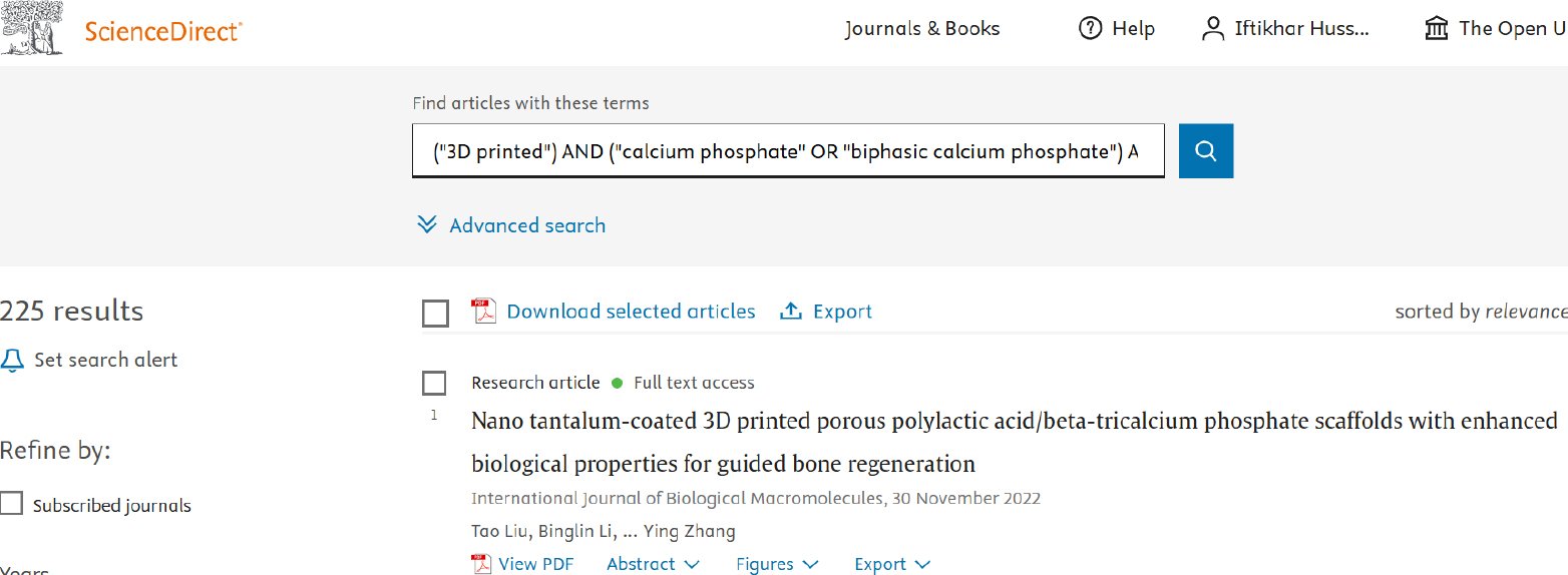
A comprehensive search was conducted in three multidisciplinary databases, PubMed, ScienceDirect, and Web of Science covering literature published from 2020 to 2025. Boolean operators and keywords were adapted to each database. A mutual search string comprised of (#D printed OR “additive manufacturing”) AND (“calcium phosphate”) AND (scaffold) AND (alveolar OR “guided bone regeneration”) AND (clinical OR randomised control trial). Searches were limited to peer-reviewed, English-language, human studies; duplicates and irrelevant records were removed during screening. The research was restricted to 2020–2025 because clinical translation of patient-specific printed calcium-phosphate (CaP) blocks is recent study as older studies reflect printing methods or materials are not comparable today. See detailed search string of each database in Appendix A.
2.3. Eligibility Criteria
2.3.1. Inclusion Criteria
Primary human clinical studies in dentistry evaluating additively manufactured (3D-printed) calcium-phosphate bioceramic scaffolds (hydroxyapatite, β-tricalcium phosphate, biphasic calcium phosphate, or calcium-phosphate cement), used for Alveolar Ridge Preservation or augmentation in maxillary/mandibular implant sites. Studies report quantitative outcomes relevant to hard-tissue regeneration e.g., CBCT ridge width/height change (mm), histomorphometry/BV/TV (%), residual graft (%), Implant Stability Quotient and Implant Stability Test (ISQ/IST), or complications with extractable data. Meta-analysis was planned if ≥2 clinically homogeneous RCT were reported with compatible outcomes. However, heterogeneity and limited reporting precluded pooling, so narrative synthesis was performed.
Other primary study designs, such as prospective and retrospective with quantitative findings, were also included in the narrative synthesis. Also, only peer-reviewed studies published in English in the years 2020-2025 were considered. The limited number of eligible studies was likely due to the relative novelty and early adoption of patient-specific 3D-printed calcium-phosphate scaffolds.
2.3.2. Exclusion Criteria
Non-primary publications (systematic reviews, meta-analyses, editorials and letters), protocols without results, animal studies and reports outside the domain of dental alveolar applications were excluded in this study.
Moreover, additive manufacturing (non-printed blocks) blocks, made of polymer material (no calcium phosphate phase) or material other than calcium phosphate were excluded in this study.
Furthermore, records with no outcomes, non-English publications, conference abstracts without full text and indications other than alveolar ridge preservation/augmentation (e.g., craniofacial defects outside the dentoalveolar ridge) were also excluded and are available in Appendices.
2.4. Data Screening
A total of 409 records were identified through database searching (PubMed = 103; ScienceDirect = 225; Web of Science = 81). After removal of 106 duplicate records, 303 titles and abstracts were screened. Of these, 158 records were excluded for being outside the scope (n = 43), involving alveolar regeneration as a secondary outcome (n = 57), publications such as reviews or conference papers (n = 25), study design filters applied for exclusion (review) (n = 26) or comprising abstract-only records (n = 7). One hundred forty-five reports were sought for full-text retrieval; 21 could not be retrieved due to missing full text. Consequently, 124 reports were assessed for eligibility, of which 117 were excluded, primarily because they were non-3D-printed or non-calcium-phosphate scaffolds (n = 87) or lacked extractable outcome data (n = 30). Seven studies met the inclusion criteria and were included in the final synthesis (Fig. 1).
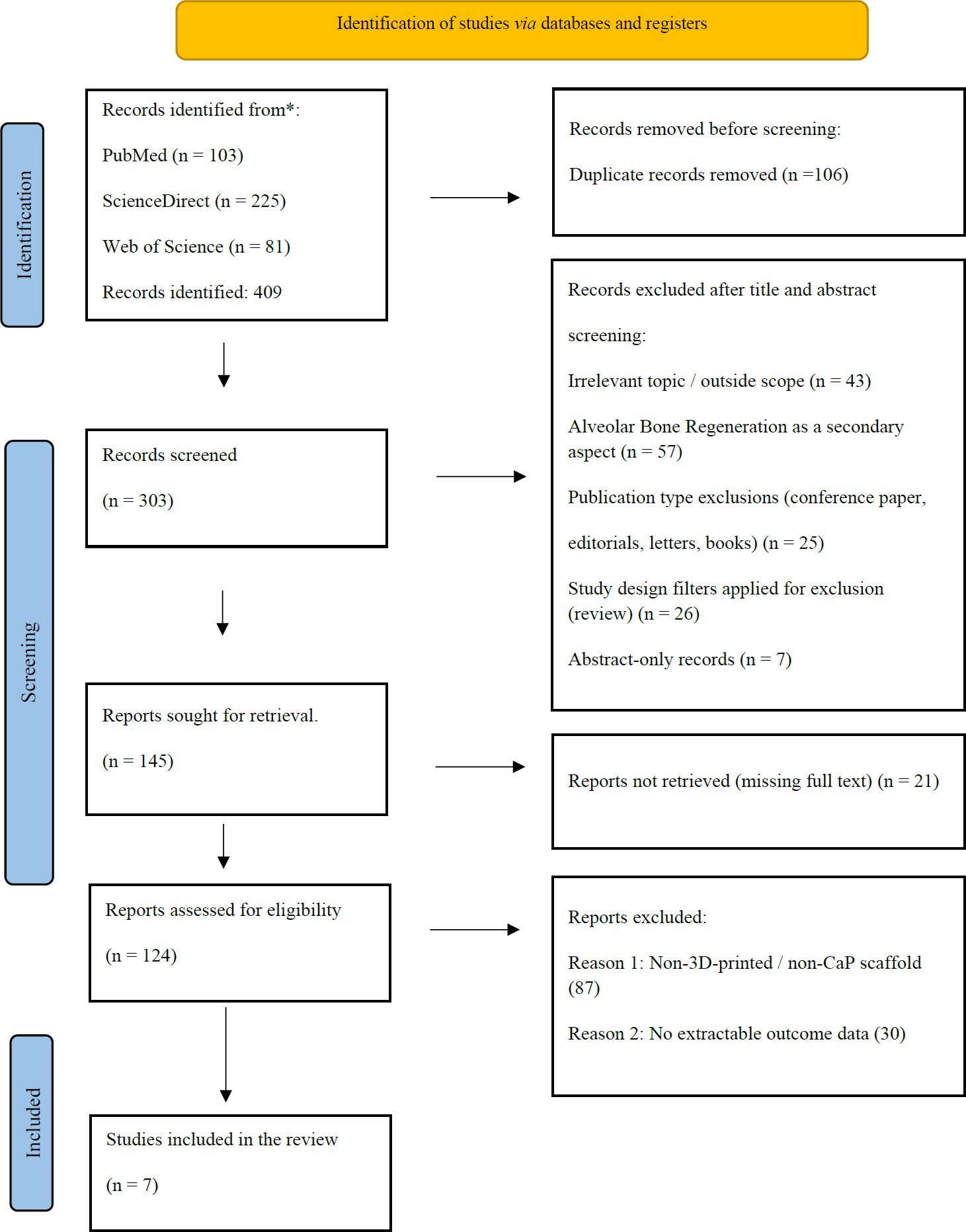
Fig. (1). PRISMA flowchart.
2.5. Data Extraction and Analysis
This review evaluated patient-specific 3D-printed calcium-phosphate scaffolds as standalone grafts versus conventional materials for dento-alveolar bone regeneration. Authors, year, aim of the study, methodology, treatment / intervention, and the findings of the study were used as variables variables shown in (Table 1). Data were extracted independently by two reviewers, with discrepancies resolved by discussion; sources and the page/figure numbers were noted for those values for which there was consensus. The provided studies were summarized into three themes: clinical effectiveness of 3D-printed calcium-phosphate scaffolds in dento-alveolar regeneration, precision-fit and workflow feasibility of patient-specific CaP scaffolds, and from CaP sources to CaP-polymer composite, material composition and manufacturing routes. Digital workflow information such as CAD/CAM software, printing and sterilising techniques were also obtained were available.
Table 1. Summary table.
| Authors | Year | Aim of the Study | Methodology | Treatment / Intervention | Findings of the Study |
| Anderson et al. [18] | 2022 | Develop a clinical-scale CBCT→CAD→print workflow for personalised CaP scaffolds and verify mechanics/biocompatibility/fit. | Bench testing (compressive strength, pH, cytotoxicity) + digital workflow validation with CBCT-derived, patient-specific prints. | OsteoInk™ (HA/α-TCP) printed scaffolds. | Stable pH (~7.2), ISO-compliant cytocompatibility, higher compressive strength vs hybrid CaP, and ~0.27 mm mean morphological deviation (precision fit). |
| Ghezzi et al. [19] | 2024 | Optimise solvent-free FDM PCL/β-TCP composites resembling cancellous bone to enhance osteogenesis. | Fabricated filaments with 40–70% β-TCP; evaluated mechanics, wettability/roughness, and cell assays. | PCL/β-TCP (up to 70%) composite scaffolds via FDM. | Higher hydrophilicity/roughness, significantly increased Young’s modulus, and improved osteoblastic growth/differentiation vs PCL. |
| Kijartorn et al. [13] | 2022 | Compare 3D-printed nano-porous HA with nanocrystalline alloplast for ARP. | Parallel RCT (n = 30); CBCT & IOS ridge dimensions, histology (%NB/%residual), implant stability (IST) at ~4 months. | 3D-printed nano-porous HA vs NanoBone® particulates under membrane. | There are no significant differences across CBCT, histology, or IST; 3DP-HA is a viable ARP alternative (non-inferior). |
| Kim et al. [12] | 2024 | Test the clinical effectiveness of customised 3D-printed alloplastic blocks vs conventional blocks in GBR. | Two-centre RCT (n = 60); micro-CT (BV/TV, TS, etc.), histology at ~5 months; clinical follow-up to rehabilitation. | Patient-customised 3D-printed BCP blocks vs conventional block grafts. | Significantly higher BV/TV and lower TS were in the customised group; other micro-CT indices were similar, and workflow convenience was noted. |
| Li et al. [20] | 2025 | Create 3D-printable PLA filaments reinforced with HA/β-TCP and assess in-vitro osteogenicity. | Thermal vs THF processing; FTIR/XRD/SEM, compression; MC3T3 proliferation, ALP, mineralisation. | PLA + HA/β-TCP (7:3) composite filaments (H-MIX, T-MIX). | Compressive strength and osteogenic markers were higher in composites; T-MIX performed best across ALP and calcium deposition. |
| Mekcha et al. [17] | 2022 | Evaluate customised 3D-printed nano-HA blocks for primary augmentation before implants. | Prospective case series (n = 12); CBCT/STL superimposition, volumetry, ISQ, histology at 6 months. | Patient-specific 3D-printed nano-HA blocks (often with CGF/PRF), membrane fixation. | Success in 10/12; horizontal gain ~3.06–3.56 mm; volume gain ~230 mm³; ISQ ~65; histology ~28.6% NB, ~19.8% residual graft; minimal trimming. |
| Zheng et al. [21] | 2025 | Verify tooth-derived CaP DLP printing feasibility and optimise process/design for alveolar Scaffolds. | DLP slurry optimisation (dispersant/solids/sintering), FEA/CFD (gyroid/diamond/hexagonal), compression, in-vitro apatite formation. | Tooth-derived CaP scaffolds via DLP; lattice comparisons. | Optimal slurry 50 vol% solids/2 wt% dispersant; best sintering ~1200 °C; gyroid showed favourable transport/mechanics; apatite-forming capacity validated. |
2.6. Quality Assessment
The included studies were appraised using Joanna Briggs Institute (JBI) for human research and QUIN for in-vitro work (Appendix B). JBI checklist showed that RCT of Kijartorn et al. 2022 [13] and Kim et al. 2024 [12] had true randomisation reported; allocation concealment and blinding were not reported, with consistent outcome measurement and appropriate statistics. Thus, it showed a moderate risk of bias. JBI Case series Mekcha et al. [17] revealed clear inclusion criteria and standardised measurement, reporting an adequate overall small risk. QUIN scores maximum of 24, with Anderson et al. 2022 [18] scoring 14/24, Ghezzi et al. 2024 [19] scoring 14/24, Li et al. 2025 [20] scoring 14/24, Zheng et al. 2025 [21] scoring 13/24; as strengths in specimen preparation, characterisation, and reporting. Common limitations were sample size, specimen randomisation, and blinding. Disagreements were resolved by consensus.
2.7. Ethical Consideration
This work does not involve direct contact with human participants and access to identifiable data; this work is based on studies, from publicly accessible sources and published work, that are used as raw data. Thus, the requirement for institutional ethics approval was not required. The review followed accepted reporting guidelines and practiced methodologically and consistently with citation and fidelity with the original data. Any possible conflicts of interest were documented for the purpose of interpreting the evidence base transparently.
3. RESULTS
The study highlights clinical effectiveness with evidence from human studies. Preclinical and engineering studies are also reported separately as translational context but they are not used to infer clinical efficacy.
3.1. Human Clinical Effectiveness Evidence (Systematic Synthesis)
Empirical data in three-dimensional printed calcium-phosphate (CaP) scaffolds in clinical settings are presently limited to two randomised controlled trials and one prospective case series, all of which have brief follow-up intervals. These papers enable a premature but tentative evaluation of clinical efficacy in Alveolar Ridge Preservation (ARP) and Guided Bone Regeneration (GBR) and highlight strong methodological and interpretive drawbacks.
3.1.1. Study Set, Indications, and Comparability
In the comprised human study, clinical evidence is based on two parallel-arm RCT and one prospective but not comparative case series representing fledgling clinical translation and not comparative effectiveness studies. The evidence is decisively divided in the case of ARP and GBR, whose differences are biological limitations, morph shape defect formation, and guided clinical purposes. The ARP RCT compared a group of post extraction sockets with 4 months interval to heal [13]. The GBR RCT, on the other hand, was done on the bigger horizontal defects, where the evaluation was done at around five to six months [12]. The outcomes in the case series at six months before the placement of the implant were also reported [17]. The difference in interventions was also significant. The nano-porous hydroxyapatite scaffold manufactured by 3D-printing for ARP was compared to a nanocrystalline alloplastic particulate bone graft under coverage of the membrane [13]. Customised 3D printed biphasic CaP blocks (HA: β-TCP 60:40) were tested in GBR, in comparison to conventional non-custom block grafts [12]. The series in the case used patient nano-HA blocks, frequently in combination with biologic adjuvants, including focused growth factors or platelet-rich fibrin [17]. These variations preclude cohort quantification, which enables evaluation of consistency or deviation of signals between indications.
3.1.2. Alveolar Ridge Preservation: Non-inferiority Within a Biological Ceiling
Kijartorn et al. [13] showed 3D-printed nano-porous hydroxyapatite scaffolds provide similar outcomes with 3D-nano crystalline alloplast in terms of radiography, histology, and implant stability, 4 months after transplantation. Changes in CBCT-based horizontal and vertical ridge also showed no significant difference between groups, while histomorphometric analysis showed a similar percentage of new bone and remnant graft material. The values of implant stability at place and initial follow-up were also found to be equal, showing clinically acceptable implant positions regardless of the type of graft. Compared with the Guided Bone Regeneration trial by Kim et al. [12], no observable bone-quality gain in ARP suggests an indication-specific drawback. The process of socket healing is constrained by early bundle bone resorption and vascular remodelling, restricting the capacity of graft material to preserve the size of ridges. Within the biological limit, findings by Kijartorn et al. [13], demonstrate non-inferiority rather than superiority of the intervention evaluated; however, the short follow-up, small sample size, and incomplete reporting of allocation concealment reduces the statistical significance with increase in risk for bias, limiting sensitivity to detect treatment effects.
3.1.3. Guided Bone Regeneration: A Consistent Early Bone-Quality Trend Signal Without Dimensional Superiority
Contrary to ARP, Kim et al. [12] report Guided Bone Regeneration RCT studies revealed statistically significant increase, with a consistent trend towards higher BV/TV in bone volume fraction (BV/TV) and a decrease in tissue surface in defects treated in customised 3D-printed biphasic CaP blocks compared to conventional blocks. These micro- CT observations indicated there was a higher density of the regenerate at around five to six months, but there was no substantial difference in absolute bone volume, surface to volume ratios, bone mineral density, and radiographic ridge sizes across the groups. This deviation, when directly compared to the ARP results of Kijartorn et al. [13], highlights the role of defect environment. GBR defects are larger, open, mechanically stiffened, and tolerant to the variations in bone quality, and ARP sockets are biologically capped. Notably, Kim et al. [12] lacked downstream clinical benefits because no evidence in regard to survival of implants, loss of marginal bone, and patient-reported outcome were studied. Thus, BV/TV increase must be regarded as an early bioindicator and not an indicator of excellent clinical outputs.
3.1.4. Supportive Feasibility Evidence and Contextual Contradictions
Mekcha et al. [17] outline in the prospective case series that patient-specific 3D-printed CaP blocks can be applied clinically, with horizontal ridge gains, volumetric gain, implant stability rate, and histological bone formation, after six months. Nonetheless, the initial graft exposures and later-on protocol adjustments that include biologic supplements complicate the ability to allocate results solely to the scaffold. In contrast to RCTs conducted by Kijartorn et al. [13] and Kim et al. [12], the study does not employ any control group and has confounding variables that do not allow making any causal inferences. Mekcha et al. [17] does not support or deny superiority claims when synthesised with the randomised evidence. Rather, it reinforces practicability and emphasises variability at earlier clinical adoption. The comparison of ARP equivalence [13], GBR bone-quality signal [12], and case-series heterogeneity [17] indicates that whatever benefit there might be in customised printed CaP scaffolds is indicator-specific, but it has yet to be established in controlled trials.
The clinical indications of the synthesised evidence suggest that 3D printed CaP scaffolds indicates biological activity and comparable to conventional grafting materials during the early phases of healing. Current evidence shows these scaffolds show superiority as compared to the present available grafting materials in terms of clinical and radiographic outcomes.
In ARP, the results interact homogeneous among the materials, which is in line with biological limitations. In GBR, there is some slight increase in bone-quality measures without evident dimensional advantage. These are internally consistent but clinically undeveloped signals. All the research are constrained due to small sample size, limited follow-up, disparate outcome measures, and failure to report on randomisation and blinding procedures. The endpoints vary significantly (CBCT compared to micro-CT compared to histology), which does not allow pooling and makes narrative comparison the only possible synthesis. Variability in CBCT protocols, histomorphometric definitions, follow-up periods, and implant-stage assessments substantially limited inter-study comparability. Therefore, the review presents only a narrative synthesis, while the existing evidence base can be seen as a hypothesis-generating, rather than a conclusive one.
3.2. Translational and Preclinical Context (Non-Clinical Evidence)
In addition to the few human experiments, 3D printing of calcium phosphate (CaP) has been studied through translational and preclinical studies on the manufacturability, architectural control, and material behaviour of 3D printed scaffolds. These studies lack clinical efficacy but provide the background information for design, feasibility and probable hypothesis that can be used in the future to optimise the clinical study.
3.2.1. Manufacturing Feasibility and Precision-Fit at Clinical Scale
The uniformity of technical feasibility is observed across the translational research: patient-specific CaP scaffolds can be produced on a clinically relevant scale. Anderson et al. [18] confirmed the feasibility of an end-to-end CBCT-to-print workflow, showcasing that customised CaP constructs could be fabricated with less than-millimetric variations from the planned design (mean 0.27 mm) while maintained neutral pH, ISO-compliant cytocompatibility and compressive behaviour under membranes. These findings overcome the long-standing translational hurdle in block grafting by fabricating patient specific constructs that adapt to the complicated shapes of defects.
Nevertheless, the data can be interpreted as feasibility evidence compared to the existing human trials particularly when compared to the limited data available from the human trials (Kijartorn et al. [13], Kim et al. [12]). Although precision-fit has been technically proven, there is no qualified controlled clinical study that improves the geometric fidelity for enhanced bone regeneration or superiority in implant outcomes. The proposed workflow advantages such as decreased intraoperative trimming or lower risk for graft contamination, have been inferred, and not evaluated by clinical studies. The work of, Anderson et al. [18] advances validation and manufactured feasibility with clinical effectiveness as a secondary option that requires further investigation.
3.2.2. Architecture and Material Studies Defining the Design Space
Material and architecture-oriented research goes further to define the design space of CaP scaffolds by studying the effects of composition, porosity, and fabrication tracks on mechanical behaviour and cellular responses. Zheng et al. [21] proved that it is possible to use tooth-derived CaP to generate scaffolds that have maintained hydroxyapatite phases, predictable shrinkage, and compressive properties in cancellous-bone scales through digital influence of light. The computational and experimental comparisons conducted on the lattice geometries showed that permeability and distribution of stress could differ significantly with architecture, which supports the point of view that internal design affected transport and load sharing. Equally, the works of polymer-ceramic composite by Ghezzi et al. [19] and Li et al. [20] depicted that the increment in ceramic content advances the stiffness, surface roughness and osteogenic indicators in-vitro. Such studies reveal through trade-offs; increased ceramic loading can lead to and enhance brittleness or handling tolerance. Notably, such studies do not give a causal relationship between a given architectural parameter and a patient level outcome. The lack of standardised reporting of architectural outcome data in human trials also restricts clinical translation of findings.
3.2.3. What Translational Evidence Can and Cannot Support
The translational and preclinical literature, when combined, can be used to make two defensible conclusions. Firstly, reproducible production of patient-specific CaP scaffolds with high geometric fidelity, acceptable mechanical behaviour, and cytocompatibility demonstrates technical feasibility in clinical testing (Anderson et al. [18], Zheng et al. [21]). Secondly, these studies produce testable hypotheses, which are plausible and define how scaffold composition and architecture can affect biological behaviour. The information is, however, incapable of bearing claims of clinical superiority, highly successful implantation, or beneficence to the patient. In vitro osteogenic markers, simulated transport measures and mechanical optimisation are not a replacement of controlled human evidence. Clinical outcomes are indication-dependent and multifactorial, as demonstrated by the differences between ARP equivalence (Kijartorn et al. [13]) and GBR a consistent early bone-quality trend (Kim et al. [12]). Translational evidence must be considered as scaffolding, not explanatory until architectural variables have been reported and run-in trials sufficiently powered to use them to explain clinical outcomes.
5. DISCUSSION
The study consists of only two randomised controlled trials and one case series with short follow-up of human evidence available; the rest of the evidence is of an engineering and in-vitro study. Primary and secondary endpoints were not prespecified: workflow-related measures, including trimming time, chairside efficiency, and risk of contamination, were not quantitatively assessed. They are presented as feasibility metrics instead of clinical benefits. In this review, clinical outcomes were prespecified; however, workflow measures were not quantitatively reported in the included trials.
Concerning the preservation of the alveolar ridge, the available evidence shows no examples of inferiority. 3D-printed calcium-phosphate scaffolds produce ridge dimensional maintenance and histological outcomes that are similar to modern alloplasts through the initial healing phase. The null outcome of ARP randomised controlled trial conforms to a biologically limited model wherein socket resorption is early and predictable. Graft materials have a predominant impact on the quality of the fills and not the alleviation of buccal plate remodelling. Here, an optimised printed calcium-phosphate scaffold can mimic the behaviour of state-of-the-art particulate alloplasts, though it would take a purposive adjustment of internal architecture to achieve biomimic socket biology. The ARP physiological remodelling ceiling is biologically constrained. Therefore, the 3D-printed calcium-phosphate scaffolds appear non-inferior to current alloplasts, providing similar dimensional and histological preservation and histological outcomes at early stages, which is a satisfactory outcome.
For Guided Bone Regeneration (GBR), the evidence suggests a small bone-quality signal for customised 3D-printed CaP blocks (e.g., higher BV/TV), while linear ridge gains appear broadly comparable at early follow-up. The GBR signal above converges with engineering-to-clinic feasibility work showing that patient-specific CaP blocks can be produced with sub-millimetric accuracy. Anderson et al. [18] report a complete digital workflow from CBCT segmentation to print, with printed OsteoInk™ scaffolds. It demonstrated ~0.27 mm mean morphological deviation from the design and maintained neutral culture pH with ISO-compliant cytocompatibility features that plausibly reduce intra-operative trimming, dead-space, and early exposure risk. The review findings indicate the advantages of a fit-driven workflow, with feasibility and biocompatibility of 3-D printed CaP scafolds supporting the trend for improved early bone-quality across the included study trend aligned with Anderson et al. [18], with CBCT-to-print scaffolds that minimize trimming and dead space. Mechanically, OsteoInk™ achieved higher compressive strength than a hybrid CaP formulation, reinforcing its space-maintenance role under membranes. The platform’s feasibility is consistent with the BV/TV findings by Kim et al. [12] supporting potential clinical applications. It states that if a scaffold truly “fits-and-fixes” the defect, it can prioritise biology (perfusion, stable clot, guided osteogenesis) over chairside turning, helping bone fraction even when linear width gains seem similar to controls.
Besides the works by Kim et al. [12] and Anderson et al. [18], the broader scope of the evidence used provides an additional background. The evidence in the ARP randomised controlled trial [11] supports non-inferiority of 3D printed calcium-phosphate scaffolds in the initial healing phases, but the case series [17] focuses on feasibility alone, with a learning curve in soft-tissue early complications and resulting adjustment of protocols. The in-vitro and production studies demonstrated that phase composition, sintering settings, composite formulation, and lattice structure can significantly impact mechanical characteristics and cellular reactions. This highlights that the term ‘3D -printed CaP’ is not a monolithic intervention [20, 21].
Mechanically, it is possible but not proven directly by experimentation on which this review rests, that sub-millimetric adaptation and internal architecture tune clot stability, perfusion dynamics, and early osteogenic processes in humans. It is plausible that precision-fit scaffolds may reduce micromotion and handling requirements; however, these workflow-related mechanisms were not quantitatively assessed in the clinical trials. It is an economic and biological burden that customisation can mitigate. Architectural choices then govern what happens inside the block. This explains why BV/TV can increase without dramatic linear gain if transport and marrow ingress are optimised locally [12]. Materials then set the boundary conditions, where Anderson et al. [18] document stable pH and better cytocompatibility for a sintered CaP paste (OsteoInk™) relative to a hybrid CaP, supporting a “bio-friendly” microenvironment for early cells. Mechanistically, it is plausible that precision-fit, internal porosity, and stable CaP phases interacting synergistically; however, this hypothesis has not yet been validated in controlled human trials.
Superiority claims likely require functionally graded porosity tailored to buccal plate biology and vascular supply rather than simply swapping material labels. For GBR, where defects are larger, and space maintenance is decisive, the combination of precision-fit blocks and supportive internal architecture plausibly confers incremental benefits. Although the BV/TV difference appears statistically small, its clinical relevance depends on whether it reflects a denser, more mature regenerate at the planned implant placement stage. Clinically, even small increases in regenerate density at implant placement may reduce perceived need for secondary augmentation or improve drilling stability, although this remains unproven in long-term studies. Micro-CT readouts higher showing higher BV/TV with reduced tissue surface represent improvements in bon regeneration that are associated with early implant stability [12]. However, between groups, the outcomes were similar and fewer cases required second-stage augmentation even with similar CBCT widths. In both indications, consistent ISQ/IST at re-entry is clinically reassuring, but harmonised timepoints and landmarks are needed to make placement-stage decisions comparable across trials.
Two orthogonal axes, envelope sufficiency and bone fraction/quality, should be interpreted together. CBCT width/height determines whether implants can be placed with ≥2 mm buccal bone, while BV/TV, % of new bone, and residual graft informs long-term remodelling and stability. Our corpus demonstrates that bone fraction can shift upwards even when linear dimensions are unchanged. This should be interpreted as added value from geometry/architecture (customisation) rather than contradiction. Micro-CT or histology becomes co-primary with CBCT in printed-block trials, with exposure/membrane complications reported uniformly.
Heterogeneity across indication (ARP vs GBR), endpoints (CBCT vs micro-CT vs histology), and time (~4 vs ~5–6 months) makes pooling fragile. Differences in CBCT acquisition parameters, landmark selection, and histomorphometric processing further reduce direct comparability between studies. A pragmatic core outcome set would include CBCT- based ridge measuring width at fixed levels (e.g., 1, 3 and 5 mm apical to the crest) using standardised vertical landmarks, along with a bone-quality panel (BV/TV, percentage of new bone, percentage of residual graft) assessed by histology or micro-CT. Implant stability (ISQ/IST) at placement and re-entry, and complication rates reported with uniform definitions and assessment windows must also be included. Trials must pre-register membrane type, adjuncts, and fixation, and report groups, means, Standard Deviation, and exact timepoints to enable meta-analysis. Standardised reporting of CAD/CAM software, printing parameters, scaffold post-processing, and sterilisation workflows is also necessary to improve reproducibility across centres. The limitations of follow-up limit generalisation on volumetric stability after remodelling, exposure of membranes with time, and maintaining early differences on bone-quality. Outcome measurement heterogeneity (e.g., different CBCT landmarking protocols, micro-CT panel definitions, and histological processing) makes inter-study comparison unreliable and meta-analytic pooling inappropriate, reducing clinical guidance accuracy.
Strengths of Anderson et al. [18] include a full CBCT (CAD/CAM) print pipeline demonstrated at a clinical scale, with rigorous materials characterisation (neutral pH, ISO-compliant cytocompatibility) and mechanical testing showing superior compressive behaviour versus a hybrid CaP. Crucially, fit accuracy is quantified (≈0.27 mm mean morphological deviation) and sub-millimetric bone-graft gaps are mapped, consistent with reduced trimming and contamination risk. The paper incorporates transparent and reproducible methods, that offers a realistic surgical benchmark, for practical quality assurance that translates neatly into clinical workflows and future RCT standardisation [21].
Material choices shape what reaches the clinic. Tooth-derived CaP slurries for DLP offers circular supply chain and high-resolution lattices. Zheng et al. [21] formalised a CAD-to-DLP workflow to optimise dispersion, solids loading and sintering parameters to demonstrate in-vitro apatite formation, supporting manufacturability and surface reactivity. The study also highlights uncertainties related to process control and batch consistency that are necessary regulators for scrutiny. Polymer-ceramic filaments for FFF/FDM could unlock on-demand patient-specific prints with improved handling. The study by Li et al. [20] showed that PLA reinforced with HA/β-TCP (7:3) raises compressive strength and boosts ALP/mineralisation in vitro, as a promising carrier while preserving CaP bioactivity, yet clinical benefit remains hypothetical without human data. Mapping these to our RCT identified differences in phase composition, sintering and lattice design plausibly to BV/TV parity, underscoring the need to disclose microstructural specs in clinical reports.
In terms of future aspects, future trials shall use grade versus uniform soils within the outer geometry of each patient, with the endpoint of 5-6 months with stratification as ARP/GBR. Plan non-inferiority on envelope (CBCT) and superiority on BV/TV; control membrane type, adjuncts (e.g., PRF/CGF), flap design, and fixation to isolate scaffold effects. Core is safety and PROs – exposure, pain and return to function. Economically, track OR, time, dispose rates, revisit rates and delayed implant costs. Clinically, the current evidence supports non-inferiority in ARP at early healing, while GBR shows a small bone-quality signal that requires confirmation in larger, longer, standardised trials for ARP when anatomy or workflow favours customisation. In GBR, customised printed CaP suggests a possible signal of better bone fraction and a cleaner workflow, especially when internal architecture is designed for transport, and the CaP phase is well-sintered and cytocompatible.
FUTURE IMPLICATIONS
Future implications include combining patient-specific outer fit with functionally graded internal porosity, built on well-characterised CaP phases, and testing in multicenter RCT with standardised CBCT and bone-quality endpoints, PROs, and cost-effectiveness. Future personalised regenerative workflows will also depend on low-dose CBCT technologies capable of maintaining accurate three-dimensional assessment while minimising radiation exposure. Meanwhile, select scaffolds by defect type and soft-tissue risk, and invest in digital planning/QA to reduce trimming and exposures.
In addition to biological outcomes, clinical translations such as patient-reported outcomes, surgical efficiency, and cost-effectiveness should also be considered in the future. This will ultimately help determine whether 3D-printed calcium-phosphate scaffolds can be used as viable alternatives to traditional grafts in clinical practice.
LIMITATIONS
This review’s inferences are constrained by the fragility of the randomised evidence. Only two human RCT met the criteria, and they spanned different indications (socket preservation vs. ridge augmentation), follow-up windows (~4 vs. ~5-6 months), and measurement modalities (CBCT linear envelope vs. micro-CT histomorphometry). In Kim et al. [12], the customised 3D-printed BCP arm showed higher BV/TV and lower tissue surface. Still, several other micro-CT endpoints and implant-stage readouts were indistinguishable from conventional blocks, implicitly limiting the clinical signal size available for pooling. Moreover, CBCT landmarking/thresholding, and possible host-graft mismatch can bias the effect estimates toward null or inflate variability. Parity reported in the ARP setting by Kijartorn et al. [13] underscores superior outcomes to demonstrate when the biologic envelope is constrained, and architecture is not explicitly optimised.
Moreover, non-randomised clinical series, although practical for feasibility, but they introduce confounding; Mekcha et al. [17] modified the protocol mid-study by adding CGF/PRF after early mucosal perforations with HA-only blocks; the subsequent reduction in complications and acceptable ISQ at 6 months is encouraging but not attributable solely to the printed scaffold. This adaptation complicates external validity and precludes inclusion in pooled RCT estimates.
Another limitation is that the architectural and materials insights are largely preclinical or computational, and materials pipelines could influence handling and degradation of tooth-derived CaP slurries optimised for DLP or PLA–HA/β-TCP filaments validated for in silico/in vitro studies on compression benches, and not in humans. Hence, any workflow advantages or safety signals inferred are provisional. Substantial heterogeneity in CBCT acquisition protocols, landmarking approaches, histological processing, and micro-CT definitions limited reproducibility and reduced confidence in cross-study comparisons
Furthermore, reporting heterogeneity limits quantitative synthesis. Across trials included in the study, not all outcomes were reported with group means, standard deviation, and sample sizes at common timepoints; histology domains (% NB, % residual graft), micro-CT metrics (BV/TV, BS/TV), and CBCT landmarks were variably defined, obliging outcome-specific pooling or narrative synthesis and raising small-study risk. Kim et al. [12] additionally noted practical uncertainties (defect segmentation, graft-site mismatch) that likely vary by centre and team expertise. Concealment and outcome assessor blinding were not consistently explicit as exposure/infection definitions and membrane strategies differed; and PROs, health-economic endpoints, and long-term (>12 mo) remodelling were rarely captured with in-vitro tests based on cytotoxicity, gene expression analysis and morphological characteristics without animal and/or human validation. Static mechanics and surface properties were emphasised in testing. The degradation kinetics, fatigue under physiological loading, sterilization effects and clinical handling were not evaluated. Therefore, findings in Ghezzi et al. [19] cannot be generalised to patient-specific architectures or clinical outcomes without human trials. Together, these constraints argue for cautious interpretation of pooled effect sizes and emphasise the need for standardised, CONSORT-compliant reporting in future RCT that co-specify architecture, fixation, and membrane protocols. Hence, limited reporting of long-term implant survival, marginal bone stability, and patient-reported outcomes was observed among included studies.
CONCLUSION
3D-printed calcium-phosphate scaffolds are a credible option for alveolar regeneration. Across randomised evidence, they are non-inferior to advanced alloplasts in Alveolar Ridge Preservation and deliver early, statistically significant BV/TV increase, demonstrating a consistent early trend towards improved bone-quality metrics in GBR settings. The signal appears to derive from precision fit, stable CaP chemistry, and architecture that supports perfusion, rather than expansion of the radiographic envelope. For practice, patient-specific blocks can streamline workflows and may reduce secondary augmentation. Head-to-head trials are required that randomise graded versus uniform porosity, use standardised outcomes, incorporate patient and economic endpoints, and extend follow-up.
LIST OF ABBREVIATIONS
ARP | = | Alveolar Ridge Preservation |
BV/TV | = | Bone Volume/Total Volume |
CAD | = | Computer-Aided Design |
CAM | = | Computer-Aided Manufacture |
CaP | = | Calcium-Phosphate |
CBCT | = | Cone Beam Computed Tomography |
GBD | = | Global Burden of Disease |
GBR | = | Guided Bone Regeneration |
HA | = | Hydroxyapatite |
ISQ | = | Implant Stability Quotient |
IST | = | Implant Stability Test |
JBI | = | Joanna Briggs Institute |
RCT | = | Randomised Controlled Trial |
SLR | = | Systematic Literature Review |
TCP | = | Tricalciumphosphate |
β-TCP | = | β-Tricalcium Phosphate |
AUTHORS’ CONTRIBUTIONS
M.S.K. was responsible for study design and data collection. W.A. and M.S.K. analysed and interpretated the data, I.U. and M.S.K. were responsible for manuscript writing and S.K. finalized the manuscript.
REPORTING GUIDELINES
PRISMA guidelines were followed in this study.
AVAILABILITY OF DATA AND MATERIALS
The data will be made available on reasonable request by contacting the corresponding author [M.S.K.].
FUNDING
None.
CONFLICT OF INTEREST
The authors declare no conflict of interest.
ACKNOWLEDGEMENTS
Declared none.
DECLARATION OF AI
None was used in the course of this research.
APPENDICES
Appendix A: Databases Check


Database | Search String (Boolean Logic) | Records Identified (n) |
PubMed | (“3D printing” OR “three-dimensional printing”) AND (“bioceramics” OR “calcium phosphate” OR hydroxyapatite OR “biphasic calcium phosphate”) AND (“alveolar ridge augmentation” OR “tooth socket” OR “ridge preservation”) AND (clinical OR human OR randomized) | 103 |
ScienceDirect | (“3D printed”) AND (“calcium phosphate” OR “biphasic calcium phosphate”) AND scaffold* AND (“guided bone regeneration”) AND implant* AND (clinical OR randomized) | 225 |
Web of Science | (“3D printed” OR “additive manufacturing”) AND (“calcium phosphate” OR hydroxyapatite OR “biphasic calcium phosphate”) AND (scaffold* OR block*) AND (alveolar OR “ridge preservation” OR “guided bone regeneration” OR GBR OR ARP) AND (dental OR implant*) AND (clinical OR randomized OR trial OR human*) | 81 |
Total | – | 409 |
Screening Stage | Reason for Exclusion | Records Excluded (n) |
Title and Abstract Screening (n = 303) | Irrelevant topic / outside scope | 43 |
– | Alveolar regeneration reported only as a secondary outcome | 57 |
– | Publication type (review articles, conference papers, editorials) | 26 |
– | Abstract-only records | 7 |
– | Subtotal excluded at title/abstract stage | 158 |
Full-Text Retrieval (n = 145) | Full text unavailable / missing | 21 |
Full-Text Eligibility Assessment (n = 124) | Non-3D-printed or non-calcium-phosphate scaffolds | 87 |
– | No extractable quantitative outcome data | 30 |
– | Subtotal excluded at full-text stage | 117 |
Included in Review | Studies meeting all eligibility criteria | 7 |
Appendix B: Quality Assessment
JBI Checklist
Kijartorn et al. 2022 [13]
Question | Yes | No | Unclear | N/A |
Was true randomization used for the assignment of participants to treatment groups? | ☑ | ☐ | ☐ | ☐ |
Was allocation to treatment groups concealed? | ☐ | ☑ | ☐ | ☐ |
Were the treatment groups similar at the baseline? | ☑ | ☐ | ☐ | ☐ |
Were participants blind to treatment assignment? | ☐ | ☐ | ☑ | ☐ |
Were those delivering treatment blind to treatment assignment? | ☐ | ☑ | ☐ | ☐ |
Were outcomes assessors blind to treatment assignment? | ☐ | ☐ | ☑ | ☐ |
Were the treatment groups treated identically, other than the intervention of interest? | ☑ | ☐ | ☐ | ☐ |
Was the follow-up complete and, if not, were the differences between groups adequately described/analysed? | ☑ | ☐ | ☐ | ☐ |
Were participants analysed in the groups to which they were randomized? | ☑ | ☐ | ☐ | ☐ |
Were outcomes measured in the same way for treatment groups? | ☑ | ☐ | ☐ | ☐ |
Were outcomes measured in a reliable way? | ☑ | ☐ | ☐ | ☐ |
Was an appropriate statistical analysis used? | ☑ | ☐ | ☐ | ☐ |
Was the trial design appropriate, and were any deviations accounted for? | ☑ | ☐ | ☐ | ☐ |
Kim et al. 2024 [12]
Question | Yes | No | Unclear | N/A |
Was true randomization used for the assignment of participants to treatment groups? | ☑ | ☐ | ☐ | ☐ |
Was allocation to treatment groups concealed? | ☑ | ☐ | ☑ | ☐ |
Were the treatment groups similar at the baseline? | ☑ | ☐ | ☐ | ☐ |
Were participants blind to treatment assignment? | ☐ | ☐ | ☑ | ☐ |
Were those delivering treatment blind to treatment assignment? | ☐ | ☑ | ☐ | ☐ |
Were outcomes assessors blind to treatment assignment? | ☐ | ☑ | ☐ | ☐ |
Were the treatment groups treated identically, other than the intervention of interest? | ☑ | ☐ | ☐ | ☐ |
Was the follow-up complete and, if not, were the differences between groups adequately described/analysed? | ☑ | ☐ | ☐ | ☐ |
Were participants analysed in the groups to which they were randomized? | ☑ | ☐ | ☐ | ☐ |
Were outcomes measured in the same way for treatment groups? | ☑ | ☐ | ☐ | ☐ |
Were outcomes measured in a reliable way? | ☑ | ☐ | ☐ | ☐ |
Was an appropriate statistical analysis used? | ☑ | ☐ | ☐ | ☐ |
Was the trial design appropriate, and were any deviations accounted for? | ☑ | ☐ | ☐ | ☐ |
Mekcha et al. 2022 [17]
Question | Yes | No | Unclear | N/A |
Were clear criteria for inclusion stated? | ☑ | ☐ | ☐ | ☐ |
Was the condition measured in a standard, reliable way for all participants? | ☑ | ☐ | ☐ | ☐ |
Were valid methods used for the identification of the condition? | ☑ | ☐ | ☐ | ☐ |
Did the case series include consecutive participants? | ☐ | ☐ | ☑ | ☐ |
Was there complete inclusion of participants? * | ☑ | ☐ | ☐ | ☐ |
Were participant demographics clearly reported? | ☑ | ☐ | ☐ | ☐ |
Was clinical information (defect type, co-interventions) clearly reported? | ☑ | ☐ | ☐ | ☐ |
Were outcomes or follow-up results clearly reported? | ☑ | ☐ | ☐ | ☐ |
Was the presenting site/setting described adequately? | ☑ | ☐ | ☐ | ☐ |
Was an appropriate statistical analysis used? | ☑ | ☐ | ☐ | ☐ |
QUIN- Quality Assessment Tool for In-Vitro Dental Studies.
No. | Criteria | Anderson et al. 2022 [18] | Ghezzi et al. 2024 [19] | Li et al. 2025 [20] | Zheng et al. 2025 [21] |
1 | Clearly stated aims/objectives | 2 | 2 | 2 | 2 |
2 | Detailed explanation of sample size calculation | 0 | 0 | 0 | 0 |
3 | Detailed explanation of sampling/specimen preparation | 2 | 2 | 2 | 2 |
4 | Details of comparison group(s) | 2 | 2 | 2 | 2 |
5 | Detailed explanation of methodology/protocol | 2 | 2 | 2 | 2 |
6 | Operator details (training/standardization) | 0 | 0 | 0 | 0 |
7 | Randomization of specimens | 0 | 0 | 0 | 0 |
8 | Method of measurement of outcomes clearly described/standardized | 2 | 2 | 2 | 2 |
9 | Outcome assessor details | 0 | 0 | 0 | 0 |
10 | Blinding | 0 | 0 | 0 | 0 |
11 | Statistical analysis is appropriate/reported | 2 | 2 | 2 | 1 |
12 | Presentation of results (tables/figures, mean ± SD, n) | 2 | 2 | 2 | 2 |
Total (max 24) | 14 | 14 | 14 | 13 |
REFERENCES
[1] Bernabé E, Marcenes W, Hernandez C, et al. Global, regional, and national levels and trends in burden of oral conditions from 1990 to 2017: A systematic analysis for the global burden of disease 2017 study. J Dent Res. 2020; 99(4): 362-73.
https://doi.org/10.1177/0022034520908533
[2] Couso‐Queiruga E, Stuhr S, Tattan M, Chambrone L, Avila‐Ortiz G. Post‐extraction dimensional changes: A systematic review and meta‐analysis. J Clin Periodontol. 2021; 48(1): 126-44.
https://doi.org/10.1111/jcpe.13390
[3] Avila‐Ortiz G, Chambrone L, Vignoletti F. Effect of alveolar ridge preservation interventions following tooth extraction: A systematic review and meta‐analysis. J Clin Periodontol. 2019; 46(S21): 195-223.
https://doi.org/10.1111/jcpe.13057
[4] Alterman M, Casap N. The Approach to Periimplantitis. Oral Maxillofac Surg Clin N Am. 2025; 37(1): 85-97.
https://doi.org/10.1016/j.coms.2024.08.003
[5] Starch-Jensen T, Deluiz D, Deb S, Bruun NH, Tinoco EM. Harvesting of autogenous bone graft from the ascending mandibular ramus compared with the chin region: A systematic review and meta-analysis focusing on complications and donor site morbidity. J Oral Maxillofac Res. 2020; 11(3): e1.
http://dx.doi.org/10.5037/jomr.2020.11301
[6] Rodriguez AE, Nowzari H. The long-term risks and complications of bovine-derived xenografts: A case series. J Indian Soc Periodontol. 2019; 23(5): 487-92.
http://dx.doi.org/10.4103/jisp.jisp_656_18
[7] Oryan A, Alidadi S, Moshiri A, Maffulli N. Bone regenerative medicine: Classic options, novel strategies, and future directions. J Orthop Surg Res. 2014; 9(1): 18.
https://doi.org/10.1186/1749-799x-9-18
[8] Bose S, Vahabzadeh S, Bandyopadhyay A. Bone tissue engineering using 3D printing. Mater Today. 2013; 16(12): 496-504.
https://doi.org/10.1016/J.MATTOD.2013.11.017
[9] Al Ali M. Chapter 4. Advanced Techniques in Porous Structure Design. Advanced Techniques in Porous Structure Design for Additive Manufacturing. John Wiley & Sons; 2025; pp. 1-224.
https://doi.org/10.1002/9781394312719.ch4
[10] Zhou J, See CW, Sreenivasamurthy S, Zhu D. Customised additive manufacturing in bone scaffolds-The gateway to precise bone defect treatment. Research. 2023; 6: 0239.
https://doi.org/10.34133/research.0239
[11] Cinici B, Yaba S, Kurt M, Yalcin HC, Duta L, Gunduz O. Fabrication strategies for bioceramic scaffolds in bone tissue engineering with generative design applications. Biomimetics. 2024; 9(7): 409.
https://doi.org/10.3390/biomimetics9070409
[12] Kim N-H, Yang B-E, On S-W, Kwon I-J, Ahn K-M, Lee J-H, Byun S-H. Customized three-dimensional printed ceramic bone grafts for osseous defects: A prospective randomized study. Sci Rep. 2024; 14(1): 3397.
https://doi.org/10.1038/s41598-024-53686-w
[13] Kijartorn P, Wongpairojpanich J, Thammarakcharoen F, Suwanprateeb J, Buranawat B. Clinical evaluation of 3D printed nano-porous hydroxyapatite bone graft for alveolar ridge preservation: A randomised controlled trial. J Dent Sci. 2022; 17(1):194-203.
https://doi.org/10.1016/j.jds.2021.05.003
[14] Ivanovski S, Breik O, Carluccio D, Alayan J, Staples R, Vaquette C. 3D printing for bone regeneration: Challenges and opportunities for achieving predictability. Periodontol 2000. 2023; 93(1): 358-84.
https://doi.org/10.1111/prd.12525
[15] Davidopoulou S, Batas L, Karakostas P, Tortopidis D, Barmpalexis P, Assimopoulou A, Angelopoulos C, Tsalikis L. Multidimensional 3D-printed scaffolds for ridge preservation and dental implant placement: A systematic review. Appl Sci. 2024; 14(2): 892.
https://doi.org/10.3390/app14020892
[16] Page MJ, McKenzie JE, Bossuyt PM, Boutron I, Hoffmann TC, Mulrow CD, Shamseer L, et al. The PRISMA 2020 statement: An updated guideline for reporting systematic reviews. BMJ. 2021; 372: n71.
http://dx.doi.org/10.1136/bmj.n71
[17] Mekcha P, Wongpairojpanich J, Thammarakcharoen F, Suwanprateeb J, Buranawat B. Customized 3D printed nanohydroxyapatite bone block grafts for implant sites: A case series. J Prosthodont Res. 2022; 67(2): 311-20.
https://doi.org/10.2186/jpr.JPR_D_22_00037
[18] Anderson M, Dubey N, Bogie K, Cao C, Li J, Lerchbacker J, Mendonça G, Kauffmann F, et al. Three-dimensional printing of clinical scale and personalised calcium phosphate scaffolds for alveolar bone reconstruction. Dent Mater. 2022; 38(3): 529-39.
https://doi.org/10.1016/j.dental.2021.12.141
[19] Ghezzi B, Matera B, Meglioli M, Rossi F, Duraccio D, Faga MG, Zappettini A, Macaluso GM, Lumetti S. Composite PCL scaffold with 70% β-TCP as a suitable structure for bone replacement. Int Dent J. 2024; 74(6): 1220-32.
https://doi.org/10.1016/j.identj.2024.02.013
[20] Li YT, Yang J-H, Chang Y-H, Chang H-H, Lin C-P. In vitro evaluation of novel composite bioceramic filaments for alveolar bone regeneration. J Dent Sci. 2025; 20(4): 2084-91.
https://doi.org/10.1016/j.jds.2025.07.012
[21] Zheng L, Xie B, Wang C, Chen D, Ai L, Liu H, Apicella A, Fan Y. Additively manufactured 3D-architected alveolar bone reconstruction scaffolds derived from extracted natural teeth: Computational modelling and feasibility evaluation. Virtual Physi Prototyp. 2025; 20(1): e2551085.
https://doi.org/10.1080/17452759.2025.2551085
Licensed
© 2026 Copyright by the Authors.
Licensed as an open access article using a CC BY 4.0 license.
Article Contents Author Rashid Ali Baloch1, * 1Indus University, Karachi, Pakistan Article History: Received: 25 August, 2026 Revised: 07 October,
Article Contents Author Muhammad Jawwad1, * 1NED University of Engineering and Technology, Karachi, Pakistan Article History: Received: 01 September, 2026
Article Contents Author Joseph Kofi Mensah1, * , David Kwashie Garr2 , David Mensah Awadzie3 1Department of Business Administration, Accra
Article Contents Author Cao Minh Thanh1, Cao Thai Phuong Thanh1, Tran Son Hai2, * 1Faculty of Information Technology, Saigon University,
Article Contents Author Muhammad Qasim1, * 1Jinnah Sindh Medical University, Karachi, Pakistan Article History: Received: 01 June, 2026 Revised: 18
Article Contents Author Eva Troja1, * 1Microbiology Laboratory, Profarma Sh.a Pharmaceutical Company, Tirana, Albania Article History: Received: 16 May, 2026