


















Article Contents
Article ID: CM2601111002
Views: 608Non-Coding RNAs in Cancer Disease Networks: A Systematic Review of lncRNA/miRNA Biomarker Translation and a Network-Depth Grading Framework
 DOI:
DOI:
 PDF
PDF
⬇ Downloads: 8
1University of Liverpool, Brownlow Hill, Liverpool, L69 3BX, United Kingdom
Received: 08 June, 2026
Accepted: 28 July, 2026
Revised: 07 July, 2026
Published: 10 August, 2026
Abstract:
Background: Non-Coding RNAs (ncRNAs) are increasingly proposed as network regulators, and biomarker studies, including long non-coding RNAs and microRNAs, are increasingly recognised as regulators of disease-associated molecular networks rather than independent biomarkers. A critical evaluation of circulating and extracellular vesicle-associated non-coding RNA biomarkers was conducted to assess their biological validity at the network scale.
Methods: The systematic review protocols were followed in accordance with the PRISMA principles. Primary studies published between 2017 and 2025 were searched in the electronic databases PubMed, Scopus, and ScienceDirect. The inclusion criteria were human-derived samples, diagnostic or prognostic clinical outcomes, and quantitative reporting of biomarker performance. Fifteen eligible studies were synthesised thematically and evaluated using a network-based framework assessing biological validity and translational readiness. Thematic analysis was used to synthesise 15 eligible studies.
Results: Non-coding RNA biomarkers had strong diagnostic and prognostic performance, with reported area under the curve (AUC) values ranging from 0.83 to 0.99 across different cancer types. Multimarker panels and network-informed biomarkers generally outperformed single-marker strategies and conventional clinical controls. Still, most studies were based on inferred network models that were not subject to mechanistic validation. Several barriers to clinical translation were consistently identified, including the homogeneity of the cohort, platform-dependent analytical variability, technically complex exosome-based workflows, and incomplete adjustment for potential confounding variables.
Conclusion: Non-coding RNAs demonstrate significant potential in the form of network-level disease readouts. However, their clinical implementation remains limited by insufficient mechanistic validation, lack of assay standardisation, and weaknesses in study design. The Network-Depth Grading Framework introduced in this review provides a structured method for assessing biological interpretability alongside translational readiness.
Keywords: Non-coding RNA, lncRNA, microRNA, ceRNA network, liquid biopsy, exosomes, biomarker translation, cancer diagnostics, systems biology.
1. INTRODUCTION
The non-coding RNAs (ncRNAs) constitute the majority of the human transcriptome and have proven to be key regulators of gene expression and disease pathways. The human genome is persistently transcribed, with most genomic regions generating RNA molecules that do not encode proteins but serve regulatory functions [1]. It is only a minute portion of the genome, roughly 2%, that is translated into protein, and the rest encodes non-coding RNAs that play important regulatory roles in cellular homeostasis and pathology [2]. Long non-coding RNAs (lncRNAs, > 200 nucleotides) and microRNAs (miRNAs, ~22 nucleotides) act as central post-transcriptional and epigenetic regulators that influence complex gene networks rather than single molecular targets [3]. Dysregulation of these ncRNA classes has been implicated in a wide range of diseases, including cancer, cardiovascular diseases, metabolic syndromes, and neurodegenerative diseases [4].
miRNAs primarily function by binding complementary sequences in target messenger RNAs, leading to translational repression or mRNA degradation [5]. LncRNAs, instead, have a wide range of mechanisms of action, including chromatin remodelling, transcriptional regulation, RNA sponging, and modulation of protein activity [6]. One of the most potent Paradigms is the competing endogenous RNA (ceRNA) hypothesis, in which lncRNAs serve as miRNA sponges, indirectly influencing the expression of downstream mRNAs and remodelling disease-related regulatory interactions [4]. These regulatory associations support the view that lncRNAs and miRNAs function as putative network regulators with inferred systems-level embedding rather than isolated molecular entities.
Despite growing interest in non-coding RNAs (ncRNAs) as regulators of gene expression and disease-associated pathways, their translation into clinically useful biomarkers remains challenging. As highlighted by Meijers et al. [7], biomarker development is often limited by reliance on isolated molecular associations or statistically derived signatures that lack mechanistic integration. Consequently, single ncRNA markers may inadequately capture the complexity of disease biology, while many predictive classifiers prioritise discriminatory performance without sufficient consideration of the regulatory networks in which ncRNAs operate. In contrast, Sheng et al. [8] emphasise the value of network-based approaches that model lncRNA–miRNA–mRNA interactions, providing greater biological interpretability and a stronger foundation for translational research. To evaluate this aspect systematically, the present review applies a Network-Depth Grading Framework that categorises studies according to the extent of regulatory-network integration, thereby distinguishing biological interpretability from diagnostic performance and assessing translational maturity beyond conventional accuracy metrics.
Although ncRNA dysregulation has been reported across numerous disease categories, cancer represents the most extensively investigated context for network-based biomarker discovery and clinical translation [9]. The global cancer burden remains substantial, with approximately 20 million new cases and nearly 10 million deaths reported worldwide, underscoring the need for more precise and biologically informative biomarkers [9]. Traditional protein-based biomarkers often demonstrate limitations in sensitivity, specificity, and dynamic responsiveness to disease progression. By comparison, Pardini et al. [10] note that ncRNAs exhibit tissue-specific expression patterns, high stability in biofluids, and early alterations during disease development, making them attractive candidates for minimally invasive diagnostics. Furthermore, circulating and exosome-associated ncRNAs have been detected in plasma, serum, urine, and saliva, supporting their potential clinical applicability [11].
Despite extensive discovery efforts, only a small proportion of reported ncRNA signatures have progressed beyond early-stage biomarker research [12]. Mu et al. [13] attribute this gap to persistent challenges in reproducibility, analytical validation, and cross-study standardisation. Previous reviews by Bhuvaneshwar and Gusev [14] and Kartika et al. [15] have examined translational pipelines and biomarker trends, respectively; however, neither evaluated ncRNA biomarkers through a structured network-depth perspective that integrates regulatory context with translational readiness. Although numerous reviews have summarised ncRNA biomarker discovery, few have systematically examined the relationship between regulatory-network depth and translational readiness. Existing reviews primarily focus on biomarker performance, bibliometric trends, or computational interaction prediction, while limited attention has been given to evaluating whether network-level biological evidence improves clinical translation. This unresolved gap provides the rationale for the present review.
The present review aims to evaluate ncRNA biomarker research through a network-centric perspective that considers regulatory interactions, pathway convergence, and translational readiness. Specifically, it examines how lncRNAs and miRNAs are positioned within disease-associated regulatory networks. It assesses the extent to which network-based evidence has been incorporated into biomarker development and validation. Unlike previous reviews that have primarily focused on biomarker performance or translational pipelines, this review introduces a Network-Depth Grading Framework to systematically evaluate biological interpretability alongside clinical translation potential. By synthesising evidence across predominantly oncology-focused studies, the review identifies key methodological strengths, translational barriers, and priorities for future biomarker development
2. MATERIALS AND METHODS
2.1. Research Design
This study adopted a systematic review design to critically evaluate the roles of long non-coding RNAs (lncRNAs) and microRNAs (miRNAs) in cancer-associated regulatory networks and their translational potential as clinical biomarkers. Given the methodological diversity of experimental, bioinformatics, and translational studies, a systematic synthesis was considered most appropriate [16]. The review was conducted in accordance with the PRISMA 2020 guidelines and followed a predefined protocol to enhance transparency, reproducibility, and minimise selection bias. The study-selection process comprised four sequential stages: identification, screening, eligibility assessment, and inclusion. Rather than quantitatively pooling outcomes, the review synthesised mechanistic, network-based, and translational evidence across eligible studies.
2.2. Search Strategy
A multi-database literature search was performed in PubMed, ScienceDirect, and Scopus, which revealed the relevant primary studies. These databases have been selected because they are widely discussed in molecular biology, genomics, and translational biomedical research [17]. Title, abstract, and keywords were used with Boolean operators to search for the maximum number of articles, as they are sensitive to and specific to their respective keywords and pertinence. The search strategy was intentionally broad to maximise retrieval of relevant studies. Boolean operators were used to combine key concepts related to non-coding RNAs, biomarker applications, and disease contexts. The search terms included (“non-coding RNA” OR “ncRNA” OR “long non-coding RNA” OR “lncRNA” OR “microRNA” OR “miRNA”) AND (“biomarker” OR “diagnosis” OR “diagnostic” OR “prognostic” OR “prediction”) AND (“cancer” OR “tumour” OR “neoplasm” OR “disease”) AND (“circulating RNA” OR “exosomal RNA” OR “extracellular vesicle” OR “liquid biopsy”). Database-specific adaptations were applied where necessary, and additional relevant studies were identified through manual screening of reference lists. Only English-language publications were included, and duplicate records were identified and removed across databases before screening. See detailed database searches in Appendix A.
2.3. Eligibility Criteria
2.3.1. Inclusion Criteria
Inclusion criteria include experimental, observational, or bioinformatics-based primary studies. The included studies were required to include a direct inquiry about lncRNAs and/or miRNA in a regulatory or ceRNA network and assess the diagnostic (or prognostic) or predictive biomarker potential. To maintain contemporaneity, the literature search was limited to primary literature published from 2017 to 2025. Research carried out in the context of human disease, such as cancer, cardiovascular disease, metabolism, or other complex pathologies, was accepted. Articles had to provide sufficient methodological detail and quantitative or qualitative measures of effect relevant to biomarker translation.
2.3.2. Exclusion Criteria
Studies were excluded if they were review articles, meta-analyses, editorials, commentaries, or conference abstracts. Preclinical mechanistic studies conducted exclusively in cell lines or animal models were excluded because, while valuable for pathway discovery, they do not directly inform biomarker performance, reproducibility, or clinical utility in humans. Although animal and cell-line studies are valuable for elucidating ncRNA regulatory networks and biological mechanisms, they do not adequately reflect inter-individual variability, biofluid complexity, or clinical biomarker performance. Therefore, they were excluded to maintain the review’s focus on human translational and diagnostic evidence. Consistent with contemporary biomarker‐translation guidance, translational relevance was defined by the use of human-derived samples (e.g., tissue, plasma, serum, urine, or exosomes) and the reporting of clinically meaningful endpoints, such as diagnostic accuracy, prognostic outcomes, or disease progression measures [18]. Studies that focused solely on protein-coding genes, without ncRNA components, were also excluded.
2.5. Data Extraction
A standardised data extraction framework was developed to ensure uniformity across studies. The extracted variables included author information, publication years, disease status, study design, type of biological sample, ncRNA type under investigation, type of network analysis, validation type, and indices of reported biomarker performance. Mechanistic information and stated translational constraints were also noted. Data extraction was independently conducted by two reviewers, with discrepancies resolved through discussion to ensure accuracy and consistency. Inter-reviewer agreement was assessed using consensus-based reconciliation rather than formal kappa statistics, given the qualitative synthesis. A predefined coding framework was iteratively refined during extraction, with themes validated through cross-reviewer comparison and discussion. Purely computational or theoretical studies were excluded unless bioinformatic analyses were combined with human clinical datasets used for biomarker evaluation.
2.6. Data Analysis
A narrative and thematic synthesis approach was chosen, considering the diversity of different methodologies and outcomes. Studies were classified by disease category and network framework, and cross-study comparisons assessed the recurrence of regulatory patterns, translational strengths, and methodological gaps. Themes were inductively identified through iterative coding of recurrent regulatory patterns, biomarker performance features, and translational limitations across studies.
2.7. Network-Depth Grading Criteria
To improve methodological rigour and reproducibility, we operationalised the network-depth grading system with a formal point-based rubric, in which each study is graded on criteria such as biomarker association, target prediction, pathway enrichment, cross-platform convergence, ceRNA network modelling, and experimental perturbation. Studies were categorised as descriptive (1 point), inferred (2-3 points), or mechanistically contextualised (≥4 points). Inter-reviewer agreement for network-depth grading was assessed using Cohen’s kappa (κ = 0.82) following independent scoring and reconciliation of all 15 studies. This framework has yet to be externally validated. Still, it has been expressly calibrated against existing translational reporting guidelines, such as REMARK (prognostic biomarker studies), TRIPOD (prediction modelling), and STARD (diagnostic accuracy), to ensure alignment with best practice in biomarker validation. Benchmarking against established reporting frameworks supports the conceptual validity and translational relevance of the grading system (Appendix B). In future work, this grading system must be formally tested on independent datasets and against multi-expert consensus.
2.8. Data Synthesis
Due to substantial heterogeneity in cancer types, specimen sources, assay platforms, biomarker panels, and validation approaches, a formal pooled diagnostic meta-analysis was not performed. Incomplete reporting of diagnostic thresholds further limited quantitative synthesis. Instead, a systematic quantitative mapping approach was adopted to summarise performance ranges, compare validation structures, and evaluate network-depth characteristics across studies while avoiding potentially misleading pooled estimates.
2.9. Ethical Considerations
The review used publicly available, already published information; thus, no ethical approval was needed. Every one of the sources was referenced, and the review procedure was performed in accordance with the standards of academic honesty and integrity.
3. RESULTS
3.1. Data Screening
Screening of data was performed according to the PRISMA requirements to have a clear and well-structured selection procedure (Fig. 1). The primary search of the databases included PubMed, Scopus, and ScienceDirect yielded 343 records (PubMed n = 123; Scopus n = 166; ScienceDirect n = 54). Before screening, duplicate records were eliminated, and a total of 283 records could be screened in terms of title and abstract. At the stage of title and abstract screening, 196 records were filtered out. Most of the exclusions occurred because the study was irrelevant or the study was not a ncRNA biomarker study (n = 93), because the study lacked a translational or biomarker focus aspect (n = 47), and journal articles type because the study was a review, editorial, commentary, conference paper, abstract or a book (n = 25), design reasons because the study failed to meet the design criteria (n = 26), and abstract-only publications (n = 5). After the screening, 87 reports were requested to retrieve them in full-text. Out of these, 23 reports were not retrieved, such as records which had unavailable full texts (n = 5) via institutional login. Considering the lack of specific lncRNA/miRNA biomarker analyses in a translational framework, 67 of them were omitted. Lastly, 15 studies were incorporated in the final qualitative synthesis because they met all the inclusion criteria.
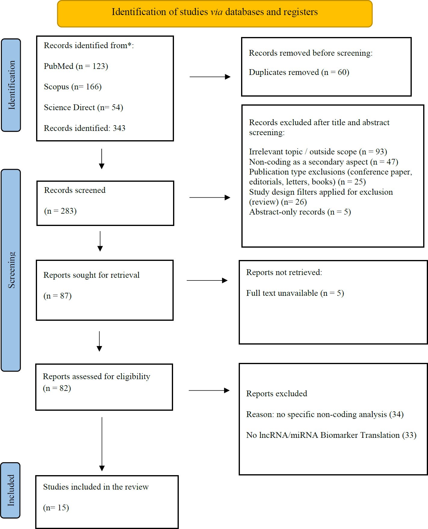
Fig. (1). PRISMA flowchart. The figure presents the process of record identification, duplicate removal, screening, full-text eligibility assessment, and final study inclusion in accordance with PRISMA 2020 guidelines. 15 studies satisfied the eligibility criteria and were included in the review.
3.2. Quality Assessment
Quality assessment revealed that the overall evidence base demonstrated low risk of bias across measurement and analytical domains, with recurring and systematic limitations primarily related to confounding control, study design, and reporting transparency (Appendix B). Prognostic studies were evaluated using the Quality in Prognostic Studies (QUIPS) tool, diagnostic studies using QUADAS-2, and prediction model studies using PROBAST. Using QUIPS, Wang et al. [19] and Zhao et al. [20] exhibited low risk of bias in prognostic factor measurement and outcome assessment, as lncRNA expression was quantified using standardised RNA-seq and qRT-PCR pipelines and survival outcomes were clearly defined and analysed using Kaplan–Meier and Cox proportional hazards models. However, a consistent pattern of moderate bias due to incomplete adjustment for clinical confounders, including tumour stage and treatment variables, was observed, reflecting common limitations of retrospective TCGA/GEO-based analyses.
Similarly, Kim et al. [21] demonstrated low risk in participant selection, biomarker measurement, and outcome definition, with support from validated protocols for exosomal isolation and quantification. Nevertheless, moderate confounding bias persisted due to limited reporting of clinicopathological covariates. Prediction modelling, assessed using PROBAST, indicated that Tao et al. [22] had a low to moderate overall risk of bias, with strong methodological performance in predictor definition, outcome ascertainment, and analytical strategy, including LASSO-based feature selection and multi-cohort external validation. However, moderate applicability concerns were identified due to population homogeneity, which may limit generalisability. Across diagnostic studies assessed with QUADAS-2, reference standards and patient flow were consistently appropriate. In contrast, case–control study designs, lack of blinding, and absence of pre-specified diagnostic thresholds represented the most recurrent sources of potential bias. Overall, the findings indicate a methodologically sound evidence base, albeit with clear opportunities to strengthen translational validity through improved adjustment for confounders, prospective designs, and enhanced reporting transparency.
3.3. Study Characteristics
The 15 studies included in this review examined the diagnostic and prognostic potential of non-coding RNAs across a range of cancer types, including non-small cell lung cancer, hepatocellular carcinoma, colorectal cancer, gastric cancer, pancreatic cancer, breast cancer, and ovarian cancer [19–24]. Most studies employed observational case-control designs, while several incorporated cohort-based validation approaches to evaluate biomarker performance in independent patient populations [23–32]. Sample sources varied across studies and included plasma, serum, whole blood, tissue specimens, and extracellular vesicles (exosomes) [19, 21, 26, 27]. The majority of investigations focused on circulating or exosomal ncRNAs as minimally invasive biomarkers. Regarding analytical methods, high-throughput discovery platforms such as RNA sequencing and microarray profiling were commonly used for candidate identification, followed by targeted validation using quantitative real-time polymerase chain reaction (qRT-PCR) or droplet digital PCR (ddPCR) [20, 23, 24].
Several studies employed multi-stage designs comprising discovery, training, and independent validation cohorts to assess biomarker reproducibility [23–25]. Bioinformatics approaches were frequently used to characterise ncRNA interactions, including ceRNA network construction, differential expression analysis, and machine-learning-based feature selection methods [19, 20, 22]. Reported outcomes included diagnostic accuracy measures, such as sensitivity, specificity, and area under the receiver operating characteristic curve (AUC), as well as prognostic endpoints including overall survival, progression-free survival, and disease recurrence [19, 22, 29]. A summary of the included studies, cancer types, sample sources, analytical platforms, and validation approaches is provided in Table 1.
Table 1. Study characteristics (Check Appendix C for complete table).
| Authors | Year | Aim of the Study | Methodology | Solution / Biomarker Identified | Findings of the Study | Outcome |
| Wang et al. | 2020 | To identify prognostic lncRNAs in NSCLC via ceRNA network analysis | Integrated bioinformatics analysis using GEO microarray data; ceRNA network construction; GO/KEGG; RT-qPCR validation; Kaplan–Meier survival analysis | lncRNA EPB41L4A-AS1 | EPB41L4A-AS1 significantly downregulated in NSCLC tissues; high expression associated with improved OS (p < 0.05); network included 33 lncRNAs, 580 mRNAs | EPB41L4A-AS1 identified as a prognostic biomarker and regulatory hub in NSCLC |
| Lin et al. | 2018 | To evaluate plasma lncRNAs for early lung cancer detection | ddPCR-based quantification of 26 lncRNAs; development and validation cohorts; ROC analysis | Plasma lncRNA signature (SNHG1, RMRP) | Combined lncRNA panel achieved 84.1% sensitivity and 87.9% specificity; independent of stage, age, sex (p > 0.05) | Plasma lncRNA signature viable for early non-invasive lung cancer diagnosis |
| Gao et al. | 2018 | To assess plasma lncRNA SNHG1 as a diagnostic biomarker for HCC | lncRNA microarray screening; plasma qRT-PCR; ROC analysis; AFP comparison | Plasma SNHG1 | SNHG1 AUC = 0.86 (HCC vs HCH); SNHG1+AFP AUC = 0.97; correlated with tumor size and TNM stage | SNHG1 superior to AFP as HCC diagnostic biomarker |
| Wang et al. | 2017 | To validate exosomal miRNAs for early-stage colon cancer detection | Plasma exosome isolation; qRT-PCR validation; ROC analysis | Exosomal miR-125a-3p | miR-125a-3p AUC = 0.685; combined with CEA AUC = 0.855; correlated with nerve infiltration (p < 0.01) | Exosomal miRNA enhances early colon cancer detection when combined with CEA |
| Kim et al. | 2023 | To identify diagnostic and prognostic exosomal miRNAs in SCLC | Serum exosome isolation; miRNA sequencing; qRT-PCR validation; ROC and survival analysis | 3-miRNA panel (miR-200b-3p, miR-3124-5p, miR-92b-5p) | Individual miRNA AUCs 0.64–0.76; combined panel AUC = 0.93; associated with poorer prognosis | Exosomal miRNA panel effective for SCLC diagnosis and prognosis |
3.4. Performance Synthesis (No Pooled Effect Estimate)
AUC values were extracted as reported in the included studies. Because study designs, diagnostic thresholds, specimen types, and validation structures were heterogeneous and many studies did not report variance estimates, central tendency was summarised descriptively using median values and ranges. No statistical weighting or pooled diagnostic accuracy estimate was calculated. The reported diagnostic AUC values (median 0.91) ranged from 0.83 to 0.99 across 15 studies. Multi-marker panels showed superior discrimination, with a median AUC of 0.93, compared with single ncRNAs, which had a median AUC of 0.88. In studies that included comparator biomarkers (e.g., AFP, CEA, CA19-9), a median incremental AUC of +0.05 to +0.12 was observed. However, 60% of studies failed to specify diagnostic thresholds, thus restricting the interpretability of sensitivity-specificity trade-offs. Network-depth stratification showed that mechanistically contextualised studies were not consistently better at demonstrating enhanced discrimination (median AUC 0.92) than inferred network studies (median AUC 0.91), suggesting that biologically motivated depth may not always translate to performance enhancement under the existing validation designs. Network depth increases biological interpretability and mechanistic plausibility, not necessarily classifier discrimination. In this dataset, AUC differences between inferred and mechanistically contextualised categories were small, indicating that translation readiness is driven more by validation architecture (cohort design, thresholds, comparators, external validation) than by network annotation depth. Studies employing retrospective case-control designs tended to report higher AUC values (median 0.94) compared with lower-risk cohort-based studies (median 0.89), suggesting possible spectrum inflation effects.
3.5. ncRNAs as Network Regulators, not Isolated Markers
A recurring conceptual distinction across the included studies concerns whether ncRNAs are investigated as isolated performance biomarkers or as regulatory components embedded within disease-associated molecular networks. To permit a systematic comparison, this review follows an analytic framework based on a network-depth framework proposed herein to achieve analytical clarity, with the studies being subdivided into three hierarchical levels: descriptive, inferred and mechanistically anchored. In descriptive studies, the associations or multi-ncRNA panels are listed, but not placed in a context of a pathway or interaction. Inferred studies do not just extend association to include target prediction, pathway enrichment or cross-platform convergence to propose regulatory embedding. Mechanistically contextualised studies have explicitly positioned ncRNAs within regulatory motifs, including ceRNA networks, and linked them to downstream molecular nodes; nevertheless, direct causal perturbation experiments have rarely been performed. In this scheme, the strongest example is Wang et al. [19], which involves EPB41L4A-AS1 as part of an lncRNA-miRNA-mRNA ceRNA network rather than as a single correlate. This plan enhances biological plausibility by embedding prognostic value within a disease-network logic.
On the other hand, most diagnostic investigations are descriptive. Kim et al. [31] report a high level of diagnostic and prognostic discrimination (AUC = 0.93) for an exosomal three-miRNA panel, but provide inadequate interpretation of the regulatory mechanisms, assay validation and clinical relationships. Yang et al. [28] focus on classification performance and only on partial translation into interpretable regulatory circuitry. With these studies, we have idealised performance-based discovery that conceives biomarkers as drivers, passengers or downstream effects within a disease network. The concept of network depth is demonstrated by Zhao et al. [20], as cross-platform consistency between the TCGA RNA-seq and GEO microarrays supports the assertion of robustness. Nonetheless, the very large AUCs observed require interpretive caution, as discrimination using enriched case-control networks and latent batch effects can be amplified and may fail to verify network control. Importantly, high AUC values alone are insufficient indicators of clinical usefulness when calibration, locked decision thresholds, and net benefit across clinically relevant risk ranges are not reported, as miscalibrated models may mislead real-world decision-making despite excellent discrimination. The work by Tao et al. [22] lies between the inferred and clinically contextualised network relevance. The fact that it is a multi-cohort study and that it assessed system-level phenotypes, including high-grade disease and progression, makes it more credible for translation. Exosome-based studies also outline the discontinuity between biological plausibility and network validation. Changes in postoperative signals and clinical utility framing are demonstrated by Zhou et al. [29] and Zhang et al. [25], suggesting their relevance at the system level. However, this is not enough evidence to prove that ncRNAs are candidate network regulators.
3.6. Specimen Type and Platform Choice Drive Reproducibility and Performance
The concept of diagnostic and prognostic performance cannot be decoupled to acquire specimen biology and assay engineering as they collectively determine the reproducibility, scalability, and clinical feasibility. Plasma and serum cell-free lncRNA assays are the most operationally simplified workflows, which entails routine phlebotomy, followed by the extraction of the RNA preceded by the quantification of the lncRNA using qRT-PCR or ddPCR assays. Their main weakness lies in the pre-analytical variability that entails haemolysis, cellular contamination as well as delays in the processing, which may be systematic in changing Ct values and diagnostic thresholds amongst labs. Lin et al. [23] partially addressed this limitation through ddPCR-based quantification, which may improve analytical precision and reproducibility compared with conventional PCR approaches. This enhances analytical accuracy and repeatability, but comes at the cost of increased platform costs and dependence on specialised instrumentation, limiting mass application. Assays using exosomes and extra-cellular vesicles seek to capitalise on the biology of vesicle-mediated RNA protection and tumour-host communication, which ideally makes them more biological. Nevertheless, operational comparison between Zhou et al. [29] and Wang et al. [19] shows the fact that workflows with exosomes create greater technical variability, especially protocol-dependent co-isolation of non-EV particles. Zhao et al. [30] also demonstrate this translational burden as Fig. (2) depicts ultracentrifugation is followed by several characterisation steps that include transmission electron microscopy, nanoparticle sizing, and immunoblotting. Such specifications provide significant turnaround time, infrastructure, and dependency on operator compared to direct plasma RNA assays. In this way, despite the common large AUCs reported in exosomal biomarkers, their scalability and reproducibility are very method-sensitive.
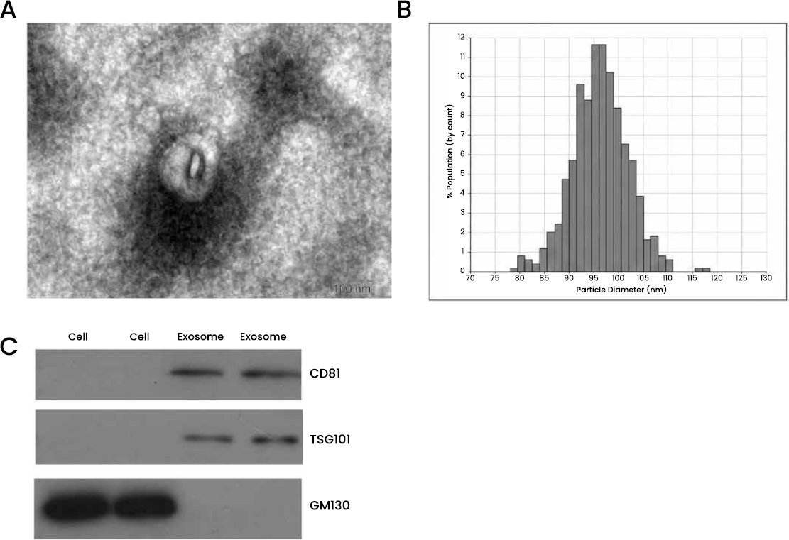
Fig. (2). Characterization of isolated exosomes; Source: Zhao et al. [30].
Urine based EV assays symbolise a viable tradeoff between biological signal and functionality. Li et al. [33] show that post-DRE urine exosomal lncRNA tests can enhance detection of prostate cancer and eliminate unnecessary biopsies, which directly relates the biomarker to clinically relevant downstream biomarker. Urine-based workflows have the advantage of non-invasive collection protocols and can be logistically simplified compared to serum EV isolation, which helps in screening and longitudinal studies through translational feasibility. Notably, the high diagnostic accuracy does not ensure the portability. An example of a hybrid integration approach is Yuan et al. [24], which a four-lncRNA plasma panel providing significant discrimination and supplemental improvement when combined with protein markers. Fig. (3) demonstrates how multi-marker lncRNA panels consistently outperform individual biomarkers, supporting the observation that integrated biomarker architectures improve diagnostic discrimination. This method balances one platform vulnerability and it would probably be simpler to standardise as compared to EV-heavy protocols. From a translational perspective, the following prioritisation of platforms can thus be realised: plasma/serum RNA assays with off-the-shelf PCR workflows, EV-based assays and lastly serum/plasma exosome-dependent pipelines. Platform choice is further contextualised through network depth. Lin et al. [23] is descriptive and presents a high level of diagnostic efficiency without regulatory interpretation. The inference of network depth is taken by Zhao et al. [20] and Yang et al. [28] to pathway and target analyses. Interestingly, causal wet-lab network validation is not demonstrated by a single experiment whose results imply that translational constraint is driven more by workflow standardisation and validation architecture than by biological discovery.
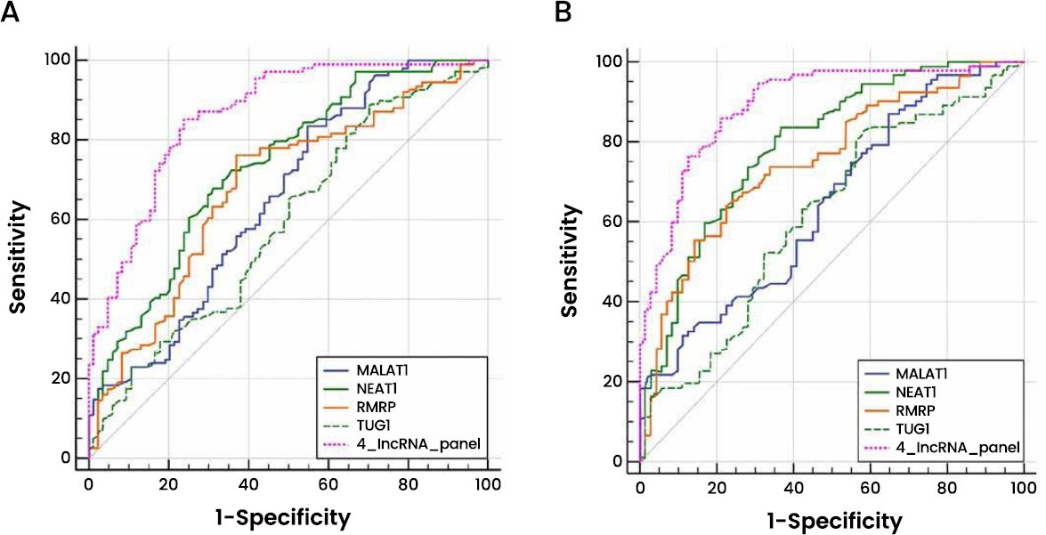
Fig. (3). Receiver operating characteristic curve analysis for the diagnosis values of the 4-lncRNA panel and each lncRNA marker. (A) Training phase. Source: Yuan et al. [24].
3.7. Translation Readiness Depends on Validation Structure, Comparators, and Generalisability
Translational preparedness may be evaluated through three interrelated dimensions: validation architecture, comparison against existing clinical standards, and evidence of integration into clinical decision pathways. Studies that address all three dimensions are more likely to demonstrate real-world applicability than those reporting diagnostic discrimination alone. From a translational perspective, decision curve analysis, calibration assessment, and net-benefit evaluation provide stronger evidence of clinical value than stand-alone measures of discrimination, particularly when biomarkers are intended to influence biopsy decisions, surveillance strategies, or treatment escalation.
Across the reviewed evidence, validation architecture emerged as a more important determinant of translation readiness than biomarker performance alone. Multi-phase designs incorporating discovery, training, and independent validation cohorts reduce optimism bias and provide an early indication of reproducibility across populations. Studies by Yuan et al. [24] and Lin et al. [23] exemplify this approach through structured development and validation phases, while Zhang et al. [25] further strengthened translational relevance by incorporating pre- and post-operative assessment and modelling clinical probability through a diagnostic nomogram (Fig. 4). Conversely, retrospectively enriched and case–control designs remain vulnerable to inflated estimates of performance and may not accurately reflect behaviour in routine clinical settings.
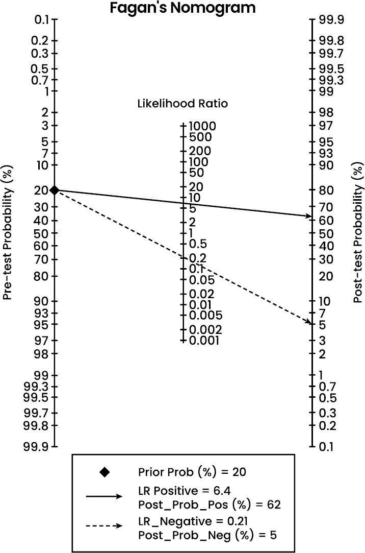
Fig. (4). Fagan’s nomogram for the calculation of the probability that an individual has gastric cancer based on the lncRNA-based diagnostic test; Source: Zhang et al. [25].
Comparator-based evaluation represents a second critical component of translational maturity. Biomarkers gain clinical relevance when they demonstrate incremental value beyond existing standards of care rather than simply achieving high levels of discrimination. Gao et al. [26] showed that plasma SNHG1 improved diagnostic performance when combined with AFP, while Zhou et al. [29] demonstrated superior performance of exosomal H19 relative to conventional markers including CA19-9, CA72-4, and CEA. As illustrated in Fig. (5), the strongest translational candidates are therefore those that complement or improve established diagnostic pathways rather than functioning as isolated molecular tests.
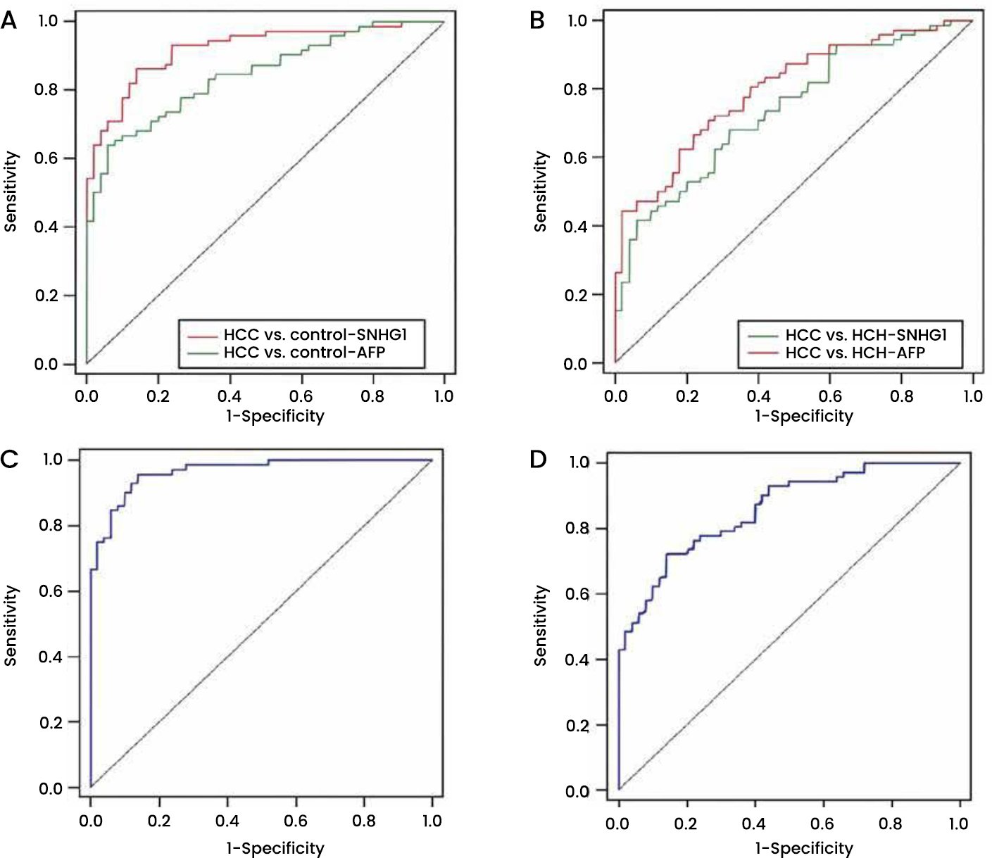
Fig. (5). Diagnostic performance of plasma SNHG1 and AFP; Sources: Gao et al. [26].
A further requirement for successful translation is evidence that biomarker outputs can influence clinical decision-making. Tao et al. [22] and Li et al. [33] moved beyond diagnostic classification by linking ncRNA signatures to biopsy triage, active-surveillance monitoring, and progression-risk stratification. Such approaches provide a clearer demonstration of potential clinical impact because they evaluate how biomarker information may alter patient management rather than simply distinguishing cases from controls.
Despite these advances, important limitations remain. Generalisability continues to be constrained by geographically restricted cohorts, incomplete reporting of diagnostic thresholds, and recurrent concerns regarding blinding and confounder control identified through QUADAS-2, QUIPS, and PROBAST assessments. These findings suggest that current barriers to implementation arise less from the biological promise of ncRNA biomarkers and more from limitations in validation design, standardisation, and clinical integration. Consequently, future studies should prioritise prospective multi-centre validation, prespecified thresholds, calibration assessment, and clinically meaningful outcome measures to strengthen the pathway from biomarker discovery to routine clinical use.
3.8. Translation Gap Across Current ncRNA Biomarker Studies
Across the included studies, four recurring translation gaps were identified. First, high diagnostic discrimination does not necessarily translate into clinical readiness, particularly when calibration, locked diagnostic thresholds, and decision-utility metrics are not reported. Second, exosome-based workflows introduce substantial technical complexity and longer turnaround times compared with direct plasma or serum assays. Third, studies employing multi-phase discovery, training, and validation architectures demonstrate stronger translational credibility than single-cohort or enriched case–control designs. Finally, most network models remain inference-based and lack experimental perturbation evidence, meaning that regulatory roles of ncRNAs are biologically plausible but not causally validated.
5. DISCUSSION
In this review, the included evidence was interpreted to assess whether circulating/EV/exosomal lncRNA and miRNA biomarkers function as network regulators and whether current study designs support translation into real-world clinical workflows. Rather than reiterating diagnostic performance, the discussion focuses on why strong discrimination alone does not guarantee clinical adoption, and where current ncRNA biomarker pipelines remain methodologically and biologically incomplete. This directly aligns with the present review’s framework integrating Zhao et al. [34], which shows that translation failure most often arises when network grounding, validation structure, and platform standardisation are insufficiently integrated.
The first critical comparison concerns mechanistic and network grounding. Candidate lncRNAs, as demonstrated by Wang et al. [32], were explicitly positioned within a ceRNA regulatory framework consistent with systems biology models of distributed gene regulation. Zhao et al. [20], Yang et al. [28], and Zhang et al. [25] assume network embedding via pathway and target methods rather than causal testing. This supports the network-depth grading applied in this review (Table 2), in which inferred and mechanistically contextualised studies offer greater interpretability than performance-only ncRNA classifiers, a point further motivated by the systems biology of Yan et al. [35]. Nevertheless, Ye et al. [36] warn that most network models remain inference-intensive, thereby enhancing plausibility but not causal control. Lu et al. [37] also demonstrate that ceRNA network construction can be prioritised by candidate hubs; however, it still requires mechanistic perturbation to establish causal biological effects.
Table 2. Network-depth grading.
| Network Depth Category | Definition | Representative Studies |
| Descriptive | Association or panel reporting without regulatory context | Lin et al. 2018 [23]; Kim et al. 2023 [21]; Yang et al. 2022 (HCC) [27] |
| Inferred | Target prediction, pathway enrichment, or cross-platform convergence | Zhao et al. 2020 [20]; Yang et al. 2022 (GBC) [28]; Zhang et al. 2017 [25] |
| Mechanistically contextualised | Explicit regulatory network modelling (no causal perturbation) | Wang et al. 2020 [19]; Tao et al. 2023 [22] |
The second comparison concerns the effects of specimen type and platform on reproducibility. Plasma-based assays reported by Lin et al. [23], Yuan et al. [24], and Gao et al. [26] demonstrate the relative analytical simplicity of cell-free ncRNA detection workflows. Trouchet et al. [38] suggests that ddPCR may improve analytical sensitivity for low-abundance circulating ncRNAs, although platform-specific limitations remain and standardisation challenges persist. This makes ddPCR-based pipelines, such as that reported by Lin et al. [23], a more viable option for clinical translation, particularly where analytical precision and assay robustness are critical requirements. Within the present framework, ddPCR strengthens analytical robustness but does not elevate network depth beyond a descriptive classification. On the other hand, Granerud et al. [39] report that ddPCR is not always the best option and should be used with caution given matrix effects, assay design, and lab workflow limitations. Importantly, network depth should be interpreted as a measure of biological interpretability rather than of diagnostic superiority, since translational maturity depends primarily on validation architecture rather than on network annotation.
Exosomes and EV biomarkers are often presented as more stable or biologically enriched than cell-free plasma RNA. This argument is partially supported by Dilsiz [40], who focuses on the importance of vesicle-mediated RNA protection and the relevance of intercellular signalling, which aligns with the extracellular communication theory. Within this review’s framework, vesicle-mediated protection is biologically plausible. Still, it does not substitute for network anchoring or translational standardisation. Nevertheless, isolation and characterisation methods introduce significant variability in product purity and yield, thereby restricting standardisation, according to the same literature. Wang et al. [32] reported that circulating exosomal miR-125a-3p shows diagnostic potential for early-stage colon cancer, supporting the biological plausibility of vesicle-associated ncRNAs as minimally invasive biomarkers,
A third comparison concerns validation architecture and generalisability. The designs used by Yuan et al. [24], Zhang et al. [25], Kim et al. [31], and Tao et al. [22] are multi-phase discovery, training, and validation. The Tao et al. [22] study is especially well validated, with clinically meaningful endpoints, as a multi-cohort study. Zhao et al. [34] state that failure to translate would be frequent when biomarkers are not reproducible across independent cohorts or populations. Lim and Lim also highlight that population diversity is critical because biomarkers can lose calibration across various clinical environments [41]. Tahir et al. [42] stress that analytical performance must be interpreted alongside workflow feasibility, a core principle of implementation science. This reinforces the review’s translation-readiness criteria, where workflow feasibility and decision-pathway integration are weighted alongside diagnostic performance. This observation is supported by Li et al. [33] and Tao et al. [22], who state that urine-based extracellular vesicle measurements provide downstream decision advantages, including the prevention of biopsy or stratification of the progression risk. Afridi et al. [43] also present arguments to the effect that biomarkers that are a part of clinical decision pathways have a higher probability of successful translation than standalone classifiers.
IMPLICATIONS FOR PRECISION ONCOLOGY
The findings of this review have broader implications for the evolving field of precision oncology, where biomarker development is increasingly shifting from single-molecule indicators toward systems-level disease characterisation. Liquid biopsy technologies, including circulating and extracellular vesicle-associated ncRNAs, offer significant opportunities for non-invasive cancer detection, longitudinal monitoring, and treatment stratification. However, the present findings suggest that diagnostic performance alone is insufficient for successful implementation, particularly when biomarkers lack biological context or robust validation. Emerging approaches in artificial intelligence and machine learning are expected to accelerate biomarker discovery by identifying complex interaction patterns across large molecular datasets, although their clinical utility remains dependent on reproducibility and interpretability. Furthermore, the integration of ncRNA signatures with multi-omics platforms, including genomics, transcriptomics, proteomics, and metabolomics, may provide a more comprehensive representation of tumour biology. Within this context, network medicine offers a valuable framework for understanding how ncRNAs interact within broader disease-associated regulatory systems, thereby supporting more biologically informed precision oncology strategies.
STRENGTHS AND LIMITATIONS
One of the strengths of the review lies in its network-based analytical framework, which goes beyond the traditional performance aggregation of ncRNAs as regulators of disease systems. The explicit grading of studies based on network depth, descriptive, inferred, and validated, is a fine and delicate appraisal of the maturity of translational biomarkers, which is not well defined in the literature on ncRNA biomarkers. This difference was particularly evident in works such as Wang et al. [19], which incorporated lncRNAs into a competing endogenous RNA (ceRNA) network, and Tao et al. [22], which linked network-sensitive classifiers to clinical progression endpoints. The strategy enables better demarcation between statistically robust but mechanistically shallow findings, such as Lin et al. [23] and Kim et al. [23], and biologically grounded biomarkers with higher translational potential [19, 25].
Methodologically, the review had stringent inclusion criteria that required human-derived samples and clinically determined outcomes, thereby maintaining translational relevance [22, 33]. The systematic identification of the bias pattern effects in heterogeneous studies was made possible by the utilisation of the existing tools of quality assessment: QUADAS-2 (studies of diagnostic accuracy) (Zhang et al. [25] and Yuan et al. [24]) and QUIPS (studies of prognostic analysis) (Wang et al. [19] and Zhou et al. [29]) as well as a prediction modelling tool (PROBAST) (Tao et al. [22]). Incorporation of these assessments enabled greater interpretative confidence and placed reported AUCs and survival associations within their methodological contexts. The clear comparison of specimen types and platforms of analysis is another positive thing, showing that translational failure is often due to workflow and standardisation failures instead of biological irrelevance (examples of the latter are given by comparing plasma ddPCR methods to workflow (Lin et al. [23]) or exosomal workflows to workflow (Kim et al. [23] and Zhou et al. [29]).
The strongest limitation of the findings is the oncology-based evidence base, in which lung and gastrointestinal cancers were overrepresented, which limits extrapolation to non-cancer disease networks where ncRNAs are also involved [19, 24, 25]. As a result, such statements about ncRNAs as candidate network regulators cannot be extended to non-cancer settings. The second weakness is the disparity in reporting standards, which greatly inhibited cross-study comparisons of measures such as cost, turnaround time, and reproducibility, which were not consistently reported [21, 27]. Thus, conclusive evidence of the implementation’s efficacy cannot be obtained. Thirdly, although network-depth grading improved conceptual clarity, classification depended, in part, on author-reported analyses, thereby introducing an element of interpretative subjectivity. A range of studies make bioinformatic inferences about regulatory engagement without experimental perturbation, which may under- or overestimate real mechanistic integration [20, 28]. Moreover, a meta-analysis was not permitted due to inconsistencies in study design, specimen type, and outcome definitions. Lastly, publication bias is still probable, with studies with high diagnostic accuracy (usually an AUC of at least 0.90) cited preferentially, overstating perceived performance among ncRNA biomarkers.
Based on the translational barriers identified across the included studies, a four-phase framework can be proposed to guide future clinical development of ncRNA biomarkers. Phase I (Analytical Validity) – At this stage, assays are standardised, reproducibility is assessed, inter-laboratory concordance is evaluated, and the pre-analytical workflow is harmonised. Phase II (Clinical Validity) entails prospective cohort validation with a predefined threshold, calibration testing, and benchmarking comparisons of 2 standard-of-care markers. Phase III (Clinical Utility) will require decision curve analysis, net reclassification improvement analysis, and health-economic modelling in representative populations. Phase IV (Implementation and Scalability) includes multi-centre implementation, cost-effectiveness analysis, regulatory fit, and real-world performance audit. The majority of the studies included in this review remain at Phase 2 case-control validation, with little advancement toward an evidence-based level of utility or implementation.
NOVEL CONTRIBUTIONS OF THE REVIEW
This review makes several novel contributions to the ncRNA biomarker literature. First, it introduces a Network-Depth Grading Framework that systematically differentiates descriptive biomarker studies from inferred and mechanistically contextualised network investigations. Second, it proposes a translation-readiness perspective that evaluates biomarkers not only by diagnostic performance but also by validation architecture, clinical comparators, and implementation potential. Third, the review integrates biological validity and clinical validity within a single analytical framework, enabling a more comprehensive assessment of biomarker maturity. Finally, it highlights the distinction between biomarker discrimination and biological interpretability, demonstrating that strong diagnostic performance does not necessarily indicate deeper mechanistic understanding or greater translational readiness.
CONCLUSION
This systematic review examined the diagnostic and prognostic capacities of lncRNA- and miRNA-based biomarker performance from a network-depth perspective, which incorporated the biological context and translational readiness. In an analysis of the 15 studies included, non-coding RNAs showed strong performance over all studies in cancer detection and prognosis, especially when combined with multi-marker signatures and network-informed biomarker models. It also showed that there was an increasing trend for the use of circulating and extracellular vesicle-associated ncRNAs as non-invasive biomarker candidates. Use of the Network-Depth Grading Framework revealed that most studies mainly investigated inferred regulatory networks, with only a few studies offering more in-depth mechanistic contextualisations. In general, the performance of biomarkers was good, but there was excellent variation among studies in terms of how mature it was in the sense of translation, depending on the information provided, study design and validation methodology used in the studies.
A key innovation of this review is the introduction of the Network-Depth Grading Framework, which provides a structured approach for differentiating descriptive biomarker studies from network-informed and mechanistically anchored investigations. Unlike conventional reviews that primarily compare diagnostic performance metrics, the framework integrates biological interpretability, validation architecture, and translational readiness. This enables a more comprehensive assessment of biomarker maturity and offers a practical roadmap for prioritising ncRNA candidates for future clinical development. In summary, the results presented foster the advancement of ncRNA markers in precision oncology as valuable clinical tools. However, available evidence currently is primarily available in oncology populations and is highly heterogeneous in methodology. The validation of the approaches, enhancement of reproducibility, and better coupling of biological and clinical evidence are expected to be future steps. A structured framework, known as the Network-Depth Grading Framework, is provided for evaluating these dimensions, and this framework could help future efforts to help close the biomarker discovery-to-clinical use gap.
LIST OF ABBREVIATIONS
AFP | = | Alpha-Fetoprotein |
AUC | = | Area Under the Curve |
CEA | = | Carcinoembryonic Antigen |
ceRNA | = | Competing Endogenous RNA |
ddPCR | = | Droplet Digital Polymerase Chain Reaction |
EV | = | Extracellular Vesicle |
GEO | = | Gene Expression Omnibus |
lncRNA | = | Long Non-Coding RNA |
miRNA | = | MicroRNA |
mpMRI | = | Multiparametric Magnetic Resonance Imaging |
ncRNA | = | Non-Coding RNA |
OS | = | Overall Survival |
PCA3 | = | Prostate Cancer Antigen 3 |
qRT-PCR | = | Quantitative Reverse Transcription Polymerase Chain Reaction |
ROC | = | Receiver Operating Characteristic |
TCGA | = | The Cancer Genome Atlas |
AUTHOR’S CONTRIBUTION
I.N. has contributed to the conceptualization of the study, development of the idea, methodology, analysis, and interpretation of the results.
CONSENT FOR PUBLICATION
Not applicable.
REPORTING GUILDLINES
PRISMA guideline has been followed.
AVAILABILITY OF DATA AND MATERIALS
All data analysed in this study are included in the published articles cited in this review.
FUNDING
No specific funding was received for the preparation of this review.
CONFLICT OF INTEREST
The authors declare no conflict of interest.
ACKNOWLEDGEMENTS
The authors acknowledge the original investigators whose work formed the basis of this review.
DECLARATION OF AI
The author used ChatGPT for language editing purposes during the preparation of this manuscript. All AI-assisted content was reviewed, verified, and approved by the author, who assumes full responsibility for the final version of the manuscript.
APPENDICES
Appendix A: Database Search

PubMed

Science Direct

Scopus
Table A2. Search strategy and study selection outcomes.
Database | Search String (Boolean Logic) | Records Identified (n) |
PubMed | “Long noncoding RNA” OR “lncRNA” AND (“microRNA” OR “miRNA”) AND (“ceRNA network” OR “molecular network”) AND (“prognostic biomarker” OR “predictive biomarker”) AND (“translational research” OR “clinical translation” OR “bench to bedside”) AND “circulating RNA” OR “exosomal RNA” AND (human disease OR cancer OR cardiovascular OR metabolic) | 123 |
Scopus | (long noncoding RNA) AND (miRNA) AND (ceRNA network) AND (prognostic marker) | 166 |
ScienceDirect | (lncRNA) AND (“microRNA”) AND (“regulatory network” OR “ceRNA network”) AND “molecular biomarker” AND (“clinical application” OR “translational medicine”) AND cell-free RNA AND pathogenesis. | 54 |
Total |
| 343 |
Table A3. PRISMA-based inclusion and exclusion summary.
Stage | PubMed | Scopus | Science Direct | Total (n) |
Records identified from databases | 123 | 166 | 54 | 343 |
Records removed before screening | – | – | – | – |
– Duplicate records removed | 22 | 29 | 9 | 60 |
Total remaining | 101 | 137 | 45 | 283 |
Records screened (title and abstract) | 101 | 137 | 45 | 283 |
Records excluded | 70 | 95 | 31 | – |
– Unrelated topic / outside scope | 33 | 45 | 15 | 93 |
– Non-coding as a secondary aspect | 17 | 23 | 7 | 47 |
– Publication type exclusions (conference paper, abstracts, editorials, letters, books) | 9 | 12 | 4 | 25 |
– Study design filters applied for exclusion (review) | 4 | 5 | 1 | 10 |
Abstract-only records | 2 | 3 | 0 | 5 |
Total excluded | 70 | 95 | 31 | 196 |
Reports sought for retrieval. | 31 | 42 | 14 | 87 |
Reports not retrieved | 2 | 2 | 1 | – |
– Full text unavailable | 2 | 2 | 1 | 5 |
Reports assessed for eligibility. | 29 | 40 | 13 | 82 |
Reports excluded (full-text assessment) | 23 | 34 | 10 | – |
– no specific non-coding analysis | 12 | 16 | 6 | 34 |
– No lncRNA/miRNA Biomarker Translation | 11 | 18 | 4 | 33 |
Studies included in the final review | 6 | 6 | 3 | 15 |
Appendix B: Quality Assessment
Table B1. QUIPS quality assessment table.
| Wang et al., 2020 | ||
| QUIPS Domain | Key Assessment Points | Risk of Bias |
| Study Participation | TCGA and GEO datasets clearly defined; NSCLC inclusion criteria reported; clinical validation cohort described | Low |
| Study Attrition | Retrospective datasets; no follow-up loss applicable; survival data completeness adequate | Low |
| Prognostic Factor Measurement | lncRNA expression quantified using standardized RNA-seq and qRT-PCR; consistent methods across samples | Low |
| Outcome Measurement | Overall survival clearly defined; standard survival endpoints used | Low |
| Study Confounding | Limited adjustment for clinical confounders; multivariate analysis partially reported | Moderate |
| Statistical Analysis & Reporting | ceRNA network construction, Kaplan–Meier, Cox regression appropriately applied | Low |
| Kim et al., 2023 | ||
| Study Participation | Clear case–control definition; SCLC patients and controls well described | Low |
| Study Attrition | Cross-sectional design; no attrition reported; complete datasets | Low |
| Prognostic Factor Measurement | Exosomal miRNAs measured using sequencing and qRT-PCR; standardized isolation protocols | Low |
| Outcome Measurement | Prognostic outcomes (survival) defined and clinically relevant | Low |
| Study Confounding | Limited reporting of treatment effects and clinical covariates | Moderate |
| Statistical Analysis & Reporting | ROC, survival analysis, and panel validation appropriately conducted | Low |
| Zhou et al., 2020 | ||
| Study Participation | Gastric cancer cohort described; inclusion/exclusion criteria stated | Low |
| Study Attrition | Observational cohort with survival follow-up; attrition handling not fully detailed | Moderate |
| Prognostic Factor Measurement | Exosomal lncRNA H19 measured using qRT-PCR; same method for all participants | Low |
| Outcome Measurement | Survival outcomes clearly defined; follow-up duration reported | Low |
| Study Confounding | Minimal adjustment for clinicopathological confounders | Moderate |
| Statistical Analysis & Reporting | Kaplan–Meier and Cox regression applied; reporting adequate | Low |
| Zhao et al., 2020 | ||
| Study Participation | TCGA (n = 216) and GEO datasets (n = 287) clearly described; NSCLC subtypes specified; inclusion based on tumor vs adjacent normal tissues | Low |
| Study Attrition | Retrospective datasets; survival data available for TCGA cohort; no loss-to-follow-up applicable | Low |
| Prognostic Factor Measurement | lncRNA expression measured using RNA-Seq (FPKM, log2) and microarray platforms; standardized normalization and feature selection applied | Low |
| Outcome Measurement | Overall survival defined; Cox regression and Kaplan–Meier analysis conducted using TCGA clinical data | Low |
| Study Confounding | Limited adjustment for clinical covariates (e.g., stage, treatment); most lncRNAs not significant after multivariable Cox analysis | Moderate |
| Statistical Analysis & Reporting | Appropriate use of differential expression analysis, machine-learning classifiers (BayesNet, Voted Perceptron), ROC, Cox regression | Low |
Table B2. PROBAST risk of bias assessment.
| Tao et al., 2023 – Urine EV lncRNA Classifier for High-Grade Prostate Cancer | ||
| Domain 1: Participants | ||
| Signalling Question | Assessment | Risk |
| Was the data source appropriate? | Multicenter cohorts (training, validation, TCGA, prospective AS cohort) clearly defined | Low |
| Were inclusion and exclusion criteria clearly specified? | Explicit criteria for GG≥2 PCa, controls, and AS cohort reported | Low |
| Were participants representative of the target population? | Patients largely from Chinese centers; limited ethnic diversity | Moderate |
| Domain 1 Overall Judgment: Moderate risk of bias | ||
| Domain 2: Predictors | ||
| Signalling Question | Assessment | Risk |
| Were predictors clearly defined and measured consistently? | Urine EV lncRNAs quantified using standardized RNA-seq and qRT-PCR | Low |
| Were predictor assessments blinded to outcome? | Molecular assays conducted independently of biopsy outcome | Low |
| Were predictors available at the time of intended use? | Urine samples collected prior to biopsy and AS outcomes | Low |
| Domain 2 Overall Judgment: Low risk of bias | ||
| Domain 3: Outcome | ||
| Signalling Question | Assessment | Risk |
| Was the outcome clearly defined? | High-grade PCa (GG≥2) and AS progression explicitly defined | Low |
| Was outcome determination blinded to predictors? | Central pathological review used for biopsy outcomes | Low |
| Was outcome assessment appropriate and reliable? | Histopathology and longitudinal AS follow-up used | Low |
| Domain 3 Overall Judgment: Low risk of bias | ||
| Domain 4: Analysis | ||
| Signalling Question | Assessment | Risk |
| Was the sample size adequate? | Large training (n = 350), validation (n = 232; n = 251), TCGA (n = 499), prospective (n = 182) | Low |
| Were predictors selected appropriately? | LASSO regression used for feature selection | Low |
| Was overfitting addressed? | Independent external validation cohorts used | Low |
| Were performance measures appropriate? | AUC, calibration plots, decision curve analysis reported | Low |
| Were missing data handled appropriately? | Minimal missing data; handling not explicitly detailed | Moderate |
| Domain 4 Overall Judgment: Low–Moderate risk of bias | ||
| Overall PROBAST Judgment | ||
| Category | Judgment | |
| Risk of Bias | Low–Moderate | |
| Concerns Regarding Applicability | Moderate (population generalisability) | |
Table B3. QUADAS-2 assessment table.
| Domain / Question | Wang et al., 2017 | Lin et al., 2018 | Zhang et al., 2017 | Yuan et al., 2020 | Zhou et al., 2020 | Li et al., 2021 | Yang et al., 2022 | Yang et al., 2022 | Kim et al., 2025 | Zhao et al., 2025 |
| Patient Selection | – | – | – | – | – | – | – | – | – | – |
| 1. Consecutive or random sample? | Unclear | Unclear | Unclear | Yes | Unclear | Yes | Unclear | Unclear | Unclear | Unclear |
| 2. Case–control design avoided? | No | No | No | No | No | No | No | No | No | No |
| 3. Inappropriate exclusions avoided? | Yes | Yes | Yes | Yes | Yes | Yes | Yes | Yes | Yes | Yes |
| Index Test | – | – | – | – | – | – | – | – | – | – |
| 4. Index test blinded to reference standard? | Unclear | Unclear | Unclear | Unclear | Unclear | Unclear | Unclear | Unclear | Unclear | Unclear |
| 5. Diagnostic threshold pre-specified? | Unclear | Unclear | Unclear | Unclear | Unclear | Unclear | Unclear | Unclear | Unclear | Unclear |
| Reference Standard | – | – | – | – | – | – | – | – | – | – |
| 6. Reference standard correctly classifies disease? | Yes | Yes | Yes | Yes | Yes | Yes | Yes | Yes | Yes | Yes |
| 7. Reference standard blinded to index test? | Unclear | Unclear | Unclear | Unclear | Unclear | Unclear | Unclear | Unclear | Unclear | Unclear |
| Flow & Timing | – | – | – | – | – | – | – | – | – | – |
| 8. Appropriate interval between tests? | Yes | Yes | Yes | Yes | Yes | Yes | Yes | Yes | Yes | Yes |
| 9. All patients received reference standard? | Yes | Yes | Yes | Yes | Yes | Yes | Yes | Yes | Yes | Yes |
| 10. Same reference standard for all? | Yes | Yes | Yes | Yes | Yes | Yes | Yes | Yes | Yes | Yes |
| 11. All patients included in analysis? | Yes | Yes | Yes | Yes | Yes | Yes | Yes | Yes | Yes | Yes |
Table B4. Summary quality assessment.
| Study (First Author, Year) | Study Type | Tool Used | Overall Risk of Bias | Main Sources of Bias Identified |
| Wang et al., 2020 | Prognostic (NSCLC) | QUIPS | Low–Moderate | Incomplete adjustment for clinical confounders |
| Kim et al., 2023 | Diagnostic/Prognostic (SCLC) | QUIPS | Low–Moderate | Limited reporting of treatment covariates |
| Zhou et al., 2020 | Diagnostic/Prognostic (GC) | QUIPS | Moderate | Attrition handling and confounder adjustment |
| Zhao et al., 2020 | Prognostic (NSCLC) | QUIPS | Low–Moderate | Retrospective design; limited multivariable adjustment |
| Tao et al., 2023 | Predictive/Prognostic (PCa) | PROBAST | Low–Moderate | Population representativeness; missing data handling |
| Wang et al., 2017 | Diagnostic (CRC) | QUADAS-2 | Moderate | Case–control design; unclear blinding |
| Lin et al., 2018 | Diagnostic (Lung cancer) | QUADAS-2 | Moderate | Case–control design; threshold not pre-specified |
| Zhang et al., 2017 | Diagnostic (GC) | QUADAS-2 | Moderate | Case–control design; unclear index test blinding |
| Yuan et al., 2020 | Diagnostic (NSCLC) | QUADAS-2 | Moderate | Case–control design; threshold specification unclear |
| Li et al., 2021 | Diagnostic (PCa) | QUADAS-2 | Low–Moderate | Case–control enrichment |
| Yang et al., 2022 (HCC) | Diagnostic (HCC) | QUADAS-2 | Moderate | Case–control design; unclear blinding |
| Yang et al., 2022 (GBC) | Diagnostic (GBC) | QUADAS-2 | Moderate | Case–control design; threshold not locked |
| Kim et al., 2025 | Diagnostic (NSCLC) | QUADAS-2 | Moderate | Case–control design; reporting transparency |
| Zhao et al., 2025 | – | – | – | – |
Table B5. Scoring table.
| Criterion | Points |
| Association-only biomarker reporting | 0 |
| Target prediction analysis | +1 |
| Pathway enrichment analysis | +1 |
| Cross-platform convergence validation | +1 |
| Explicit ceRNA network construction | +2 |
| Experimental perturbation validation | +3 |
Appendix C
| Authors | Year | Aim of the Study | Methodology | Solution / Biomarker Identified | Findings of the Study | Outcome |
| Wang et al. | 2020 | To identify prognostic lncRNAs in NSCLC via ceRNA network analysis | Integrated bioinformatics analysis using GEO microarray data; ceRNA network construction; GO/KEGG; RT-qPCR validation; Kaplan–Meier survival analysis | lncRNA EPB41L4A-AS1 | EPB41L4A-AS1 significantly downregulated in NSCLC tissues; high expression associated with improved OS (p < 0.05); network included 33 lncRNAs, 580 mRNAs | EPB41L4A-AS1 identified as a prognostic biomarker and regulatory hub in NSCLC |
| Lin et al. | 2018 | To evaluate plasma lncRNAs for early lung cancer detection | ddPCR-based quantification of 26 lncRNAs; development and validation cohorts; ROC analysis | Plasma lncRNA signature (SNHG1, RMRP) | Combined lncRNA panel achieved 84.1% sensitivity and 87.9% specificity; independent of stage, age, sex (p > 0.05) | Plasma lncRNA signature viable for early non-invasive lung cancer diagnosis |
| Gao et al. | 2018 | To assess plasma lncRNA SNHG1 as a diagnostic biomarker for HCC | lncRNA microarray screening; plasma qRT-PCR; ROC analysis; AFP comparison | Plasma SNHG1 | SNHG1 AUC = 0.86 (HCC vs HCH); SNHG1+AFP AUC = 0.97; correlated with tumor size and TNM stage | SNHG1 superior to AFP as HCC diagnostic biomarker |
| Wang et al. | 2017 | To validate exosomal miRNAs for early-stage colon cancer detection | Plasma exosome isolation; qRT-PCR validation; ROC analysis | Exosomal miR-125a-3p | miR-125a-3p AUC = 0.685; combined with CEA AUC = 0.855; correlated with nerve infiltration (p < 0.01) | Exosomal miRNA enhances early colon cancer detection when combined with CEA |
| Kim et al. | 2023 | To identify diagnostic and prognostic exosomal miRNAs in SCLC | Serum exosome isolation; miRNA sequencing; qRT-PCR validation; ROC and survival analysis | 3-miRNA panel (miR-200b-3p, miR-3124-5p, miR-92b-5p) | Individual miRNA AUCs 0.64–0.76; combined panel AUC = 0.93; associated with poorer prognosis | Exosomal miRNA panel effective for SCLC diagnosis and prognosis |
| Li et al. | 2021 | To develop urine exosomal lncRNA assay for prostate cancer detection | Multicenter retrospective study; post-DRE urine EV isolation; qRT-PCR; ROC, DCA | Urine exosomal PCA3 + MALAT1 | Combined assay AUC = 0.828 (PCa); AUC = 0.831 (clinically significant PCa); avoided 24.2% unnecessary biopsies | Urine lncRNA assay improves PCa detection and biopsy decision-making |
| Tao et al. | 2023 | To construct urine EV lncRNA classifier for high-grade PCa and progression risk | RNA-seq (TAHSY, TCGA, GEO); LASSO; multicenter validation; prospective AS cohort | 3-lncRNA classifier (Clnc) | Clnc outperformed PCA3, mpMRI, PCPT-RC; validated across cohorts (n > 1200); independent predictor of AS progression | Robust diagnostic and prognostic urine EV lncRNA classifier |
| Yuan et al. | 2020 | To identify circulating lncRNAs for NSCLC diagnosis | Four-phase case–control study (discovery, training, verification, expansion); plasma qRT-PCR; ROC analysis | 4-lncRNA panel (RMRP, NEAT1, TUG1, MALAT1) | Training AUC = 0.86; verification AUC = 0.89; sensitivity 78.9% in stage I–II; outperformed CEA/CA125/CYFRA21-1 | Circulating lncRNA panel provides robust early NSCLC diagnosis |
| Zhang et al. | 2017 | To discover novel circulating lncRNAs for gastric cancer detection | Genome-wide lncRNA microarray; multi-phase validation; logistic regression; nomogram | 5-lncRNA index (TINCR, CCAT2, AOC4P, BANCR, LINC00857) | Diagnostic AUC = 0.91 (95% CI 0.88–0.95); index decreased post-surgery (p = 0.016); superior to CEA (p < 0.001) | Plasma lncRNA index effective for GC detection and disease monitoring |
| Zhou et al. | 2020 | To assess serum exosomal lncRNA H19 in GC diagnosis and prognosis | Prospective cohort; serum exosome isolation; qRT-PCR; ROC and survival analysis | Exosomal lncRNA H19 | AUC = 0.849; significantly higher than CA19-9, CA72-4, CEA; levels decreased post-surgery (p < 0.05); correlated with TNM stage | Exosomal H19 is a diagnostic and prognostic GC biomarker |
| Yang et al. (HCC) | 2022 | To evaluate exosomal miRNA panels for HCC diagnosis | miRNA microarray; qRT-PCR validation; ROC analysis | Exosomal miRNA panel (miR-26a, miR-29c, miR-199a) | AUC = 0.994 (HCC vs healthy); sensitivity 100%, specificity 96%; superior to AFP | Exosomal miRNA panel enables high-accuracy HCC diagnosis |
| Yang et al. (GBC) | 2022 | To develop non-invasive exosomal miRNA signature for gallbladder carcinoma | Three-step discovery-training-validation; qRT-PCR; ROC; pathway analysis | 5-miRNA signature (miR-552-3p, miR-581, miR-4433a-3p, miR-496, miR-203b-3p) | Validation AUC = 0.905; sensitivity 81.4%, specificity 86.6%; superior to CA199/CEA | Exosomal miRNA signature suitable for GBC diagnosis and prognosis prediction |
| Zhao et al. | 2020 | To identify lncRNA biomarkers using integrative cross-platform analyses | TCGA + GEO datasets; machine learning; ROC validation | 8-lncRNA classifier | Training AUC = 98.5%; validation AUC up to 99.2%; consistent across RNA-seq and microarray | Multi-lncRNA classifier provides high diagnostic accuracy for lung cancer |
| Zhao et al. | 2025 | To validate serum exosomal miR-205-5p in colorectal cancer | Ultracentrifugation-isolated exosomes; qRT-PCR; clinical correlation | Exosomal miR-205-5p | Significantly downregulated in CRC (p < 0.0001); increased post-surgery (p = 0.0053); lower in early-stage CRC | miR-205-5p is a non-invasive CRC diagnostic biomarker |
| Kim et al. | 2025 | To develop a non-invasive exosomal miRNA panel for early-stage NSCLC detection | Four-phase design: discovery (NGS, n=76), validation (qPCR, n=75), optimization (UDR platform), confirmation (target-gene and enrichment analysis); serum exosome isolation and profiling | Optimised four-miRNA exosomal panel (miR-150-5p, miR-301b-3p, miR-369-3p, miR-497-5p) using UDR ratio-based scoring | The four-miRNA panel achieved AUC 0.952 in discovery and 0.933 in validation; targets linked to VEGFA, PTEN, and BCL2 pathways | Demonstrates strong diagnostic potential for early-stage NSCLC, with inferred network embedding but without wet-lab causal validation |
REFERENCES
[1] Poliseno L, Lanza M, Pandolfi PP. Coding, or non-coding, that is the question. Cell Res. 2024 Sep;34(9):609-29. https://doi.org/10.1038/s41422-024-00975-8
[2] Park EG, Ha H, Lee DH, Kim WR, Lee YJ, Bae WH, et al. Genomic analyses of non-coding RNAs overlapping transposable elements and its implication to human diseases. Int J Mol Sci. 2022 Aug 11;23(16):8950. https://doi.org/10.3390/ijms23168950
[3] Statello L, Guo CJ, Chen LL, Huarte M. Gene regulation by long non-coding RNAs and its biological functions. Nat Rev Mol Cell Biol. 2021 Feb;22(2):96-118. https://doi.org/10.1038/s41580-020-00315-9
[4] Ilieva MS. Non-coding RNAs in neurological and neuropsychiatric disorders: unraveling the hidden players in disease pathogenesis. Cells. 2024 Jun 19;13(12):1063. https://doi.org/10.3390/cells13121063
[5] Naeli P, Winter T, Hackett AP, Alboushi L, Jafarnejad SM. The intricate balance between microRNA-induced mRNA decay and translational repression. FEBS J. 2023 May;290(10):2508-24. https://doi.org/10.1111/febs.16422
[6] Kopp F, Mendell JT. Functional classification and experimental dissection of long noncoding RNAs. Cell. 2018 Jan 25;172(3):393-407. https://doi.org/10.1016/j.cell.2018.01.011
[7] Meijers WC, Bayes-Genis A, Mebazaa A, Bauersachs J, Cleland JG, Coats AJ, et al. Circulating heart failure biomarkers beyond natriuretic peptides: review from the Biomarker Study Group of the Heart Failure Association (HFA), European Society of Cardiology (ESC). Eur J Heart Fail. 2021 Oct;23(10):1610-32. https://doi.org/10.1002/ejhf.2346
[8] Sheng N, Huang L, Gao L, Cao Y, Xie X, Wang Y. A survey of computational methods and databases for lncRNA-miRNA interaction prediction. IEEE/ACM Trans Comput Biol Bioinform. 2023 Apr 4;20(5):2810-26. Available from: https://ieeexplore.ieee.org/stamp/stamp.jsp?arnumber=10091917
[9] Sung H, Ferlay J, Siegel RL, Laversanne M, Soerjomataram I, Jemal A, et al. Global cancer statistics 2020: GLOBOCAN estimates of incidence and mortality worldwide for 36 cancers in 185 countries. CA Cancer J Clin. 2021 May;71(3):209-49. https://doi.org/10.3322/caac.21660
[10] Pardini B, Sabo AA, Birolo G, Calin GA. Noncoding RNAs in extracellular fluids as cancer biomarkers: the new frontier of liquid biopsies. Cancers (Basel). 2019 Aug 14;11(8):1170. https://doi.org/10.3390/cancers11081170
[11] Ghani MU, Du L, Moqbel AQ, Zhao E, Cui H, Yang L, et al. Exosomal ncRNAs in liquid biopsy: a new paradigm for early cancer diagnosis and monitoring. Front Oncol. 2025 Jul 9; 15:1615433. https://doi.org/10.3389/fonc.2025.1615433
[12] Diamantopoulos MA, Boti MA, Sarri T, Scorilas A. Non-coding RNAs in health and disease: from biomarkers to therapeutic targets. LabMed. 2025;2(3):17. https://doi.org/10.3390/labmed2030017
[13] Mu D, Han B, Huang H, Zheng Y, Zhang J, Shi Y. Unraveling the advances of non-coding RNAs on the tumor microenvironment: innovative strategies for cancer therapies. J Transl Med. 2025 Jun 2;23(1):614. https://doi.org/10.1186/s12967-025-06629-6
[14] Bhuvaneshwar K, Gusev Y. Translational bioinformatics and data science for biomarker discovery in mental health: an analytical review. Brief Bioinform. 2024 Mar 1;25(2):bbae098. https://doi.org/10.1093/bib/bbae098
[15] Kartika AI, Dafip M, Wijayanti N, Heriyanto DS, Haryana SM, Taroeno-Hariadi KW. Research trends in microRNA profiling as a biomarker for lung adenocarcinoma via liquid biopsy: A bibliometric analysis. Narra J. 2025 Jan 16;5(1):e1372. https://doi.org/10.52225/narra.v5i1.1372
[16] Azarian M, Yu H, Shiferaw AT, Stevik TK. Do we perform systematic literature review right? A scientific mapping and methodological assessment. Logistics. 2023 Nov 27;7(4):89. https://doi.org/10.3390/logistics7040089
[17] Yeung AW. Comparison between Scopus, Web of Science, PubMed and publishers for mislabelled review papers. Curr Sci. 2019 Jun 10;116(11):1909-14. Available from: https://www.jstor.org/stable/10.2307/27138143
[18] Biomarkers of Aging Consortium. Validation of biomarkers of aging. Nat Med. 2024 Feb;30(2):360-72. https://doi.org/10.1038/s41591-023-02784-9
[19] Wang M, Zheng S, Li X, Ding Y, Zhang M, Lin L, et al. Integrated analysis of lncRNA-miRNA-mRNA ceRNA network identified lncRNA EPB41L4A-AS1 as a potential biomarker in non-small cell lung cancer. Front Genet. 2020 Sep 18; 11:511676. https://doi.org/10.3389/fgene.2020.511676
[20] Zhao T, Khadka VS, Deng Y. Identification of lncRNA biomarkers for lung cancer through integrative cross-platform data analyses. Aging (Albany NY). 2020 Jul 16;12(14):14506-27. Available from: https://pmc.ncbi.nlm.nih.gov/articles/PMC7425463/pdf/aging-12-103496.pdf
[21] Kim DH, Park H, Choi YJ, Im K, Lee CW, Kim DS, et al. Identification of exosomal microRNA panel as diagnostic and prognostic biomarker for small cell lung cancer. Biomark Res. 2023 Sep 13;11(1):80. https://doi.org/10.1186/s40364-023-00517-1
[22] Tao W, Wang BY, Luo L, Li Q, Meng ZA, Xia TL, et al. A urine extracellular vesicle lncRNA classifier for high-grade prostate cancer and increased risk of progression: A multi-center study. Cell Rep Med. 2023 Oct 17;4(10):101240. https://doi.org/10.1016/j.xcrm.2023.101240
[23] Lin Y, Leng Q, Zhan M, Jiang F. A plasma long noncoding RNA signature for early detection of lung cancer. Transl Oncol. 2018 Oct;11(5):1225-31. https://doi.org/10.1016/j.tranon.2018.07.016
[24] Yuan S, Xiang Y, Guo X, Zhang Y, Li C, Xie W, et al. Circulating long noncoding RNAs act as diagnostic biomarkers in non-small cell lung cancer. Front Oncol. 2020 Dec 7; 10:537120. https://doi.org/10.3389/fonc.2020.537120
[25] Zhang K, Shi H, Xi H, Wu X, Cui J, Gao Y, et al. Genome-wide lncRNA microarray profiling identifies novel circulating lncRNAs for detection of gastric cancer. Theranostics. 2017 Jan 1;7(1):213. https://doi.org/10.7150/thno.16044
[26] Gao S, Xu X, Wang Y, Zhang W, Wang X. Diagnostic utility of plasma lncRNA small nucleolar RNA host gene 1 in patients with hepatocellular carcinoma. Mol Med Rep. 2018 Sep;18(3):3305-13. https://doi.org/10.3892/mmr.2018.9336
[27] Yang J, Dong W, Zhang H, Zhao H, Zeng Z, Zhang F, et al. Exosomal microRNA panel as a diagnostic biomarker in patients with hepatocellular carcinoma. Front Cell Dev Biol. 2022 Sep 23; 10:927251. https://doi.org/10.3389/fcell.2022.927251
[28] Yang P, Song F, Yang X, Yan X, Huang X, Qiu Z, et al. Exosomal MicroRNA signature acts as an efficient biomarker for non-invasive diagnosis of gallbladder carcinoma. iScience. 2022 Sep 16;25(9):104938. https://doi.org/10.1016/j.isci.2022.104816
[29] Zhou H, Shen W, Zou H, Lv Q, Shao P. Circulating exosomal long non-coding RNA H19 as a potential novel diagnostic and prognostic biomarker for gastric cancer. J Int Med Res. 2020 Jul;48(7):300060520934297. http://dx.doi.org/10.1177/0300060520934297
[30] Zhao Y, Zhao Y, Liu L, Li G, Wu Y, Cui Y, et al. Tumor-exosomal miR-205-5p as a diagnostic biomarker for colorectal cancer. Clin Transl Oncol. 2025 Mar;27(3):1185-97. https://doi.org/10.1007/s12094-024-03647-6
[31] Kim YJ, Kang DH, Cho H, Chung C, Lee JE, Shin YB. Exosomal microRNA Panels for Detecting Early-Stage Non-Small Cell Lung Cancer. Diagnostics. 2025 Oct 28;15(21):2735. https://doi.org/10.3390/diagnostics15212735
[32] Wang J, Yan F, Zhao Q, Zhan F, Wang R, Wang L, et al. Circulating exosomal miR-125a-3p as a novel biomarker for early-stage colon cancer. Sci Rep. 2017 Jun 23;7(1):4150. https://doi.org/10.1038/s41598-017-04386-1
[33] Li Y, Ji J, Lyu J, Jin X, He X, Mo S, et al. A novel urine exosomal lncRNA assay to improve the detection of prostate cancer at initial biopsy: a retrospective multicenter diagnostic feasibility study. Cancers. 2021 Aug 13;13(16):4075. https://doi.org/10.3390/cancers13164075
[34] Zhao Q, Zhang C, Zhang W, Zhang S, Liu Q, Guo Y. Applications and challenges of biomarker-based predictive models in proactive health management. Front Public Health. 2025 Aug 18; 13:1633487. https://doi.org/10.3389/fpubh.2025.1633487
[35] Yan J, Risacher SL, Shen L, Saykin AJ. Network approaches to systems biology analysis of complex disease: integrative methods for multi-omics data. Brief Bioinform. 2018 Nov;19(6):1370-81. https://doi.org/10.1093/bib/bbx066
[36] Ye W, Xiang N, Wang Q, Lu Y. Role of circular RNA as competing endogenous RNA in ovarian cancer. Int J Mol Med. 2024 Mar 5;53(5):41. https://doi.org/10.3892/ijmm.2024.5365
[37] Lu F, Zeng N, Xiao X, Wang X, Gong H, Lei H. Exploring the ceRNA network involving AGAP2-AS1 as a novel biomarker for preeclampsia. Sci Rep. 2024 Nov 9;14(1):27330. https://doi.org/10.1038/s41598-024-79224-2
[38] Trouchet A, Gines G, Benhaim L, Taly V. Digital PCR: from early developments to its future application in clinics. Lab Chip. 2025;25(16):3921-61. https://doi.org/10.1039/d5lc00055f
[39] Granerud BK, Kaarbø M, Al-Baldawi H, Norwegian SARS-CoV-2 Study Group Investigators, Otterdal K, Halvorsen B, et al. Droplet digital PCR or Real-Time PCR as a method for quantifying SARS-CoV-2 RNA in Plasma-Is there a difference? Viruses. 2025 May 28;17(6):772. https://doi.org/10.3390/v17060772
[40] Dilsiz N. A comprehensive review on recent advances in exosome isolation and characterization: Toward clinical applications. Transl Oncol. 2024 Dec 1;50:102121. https://doi.org/10.1016/j.tranon.2024.102121
[41] Lim EC, Lim CE. Translational potential of RNA biomarkers in mental health: a narrative review. NeuroMarkers. 2025 Sep 24:100127. https://doi.org/10.1016/j.neumar.2025.100127
[42] Tahir MF, Ishtiaq S, Anas M, Irfan A, Jaremko M, Ercişli S, et al. Integrating biotechnology into diagnostic labs: Innovations in PCR and molecular procedures. Curr Res Biotechnol. 2025 Aug 6;100325. https://doi.org/10.1016/j.crbiot.2025.100325
[43] Afridi WA, Strachan S, Kasetsirikul S, Pannu AS, Soda N, Gough D, et al. Potential avenues for exosomal isolation and detection methods to enhance small-cell lung cancer analysis. ACS Meas Sci Au. 2023 Feb 14;3(3):143-61. https://doi.org/10.1021/acsmeasuresciau.2c00068
Licensed
© 2026 Copyright by the Authors.
Licensed as an open access article using a CC BY 4.0 license.
Article Contents Author Joseph Kofi Mensah1, * , David Kwashie Garr2 , David Mensah Awadzie3 1Department of Business Administration, Accra
Article Contents Author Cao Minh Thanh1, Cao Thai Phuong Thanh1, Cao Minh Thanh2, * 1Faculty of Information Technology, Saigon University,
Article Contents Author Muhammad Qasim1, * 1Jinnah Sindh Medical University, Karachi, Pakistan Article History: Received: 01 June, 2026 Accepted:
Article Contents Author Eva Troja1, * 1Microbiology Laboratory, Profarma Sh.a Pharmaceutical Company, Tirana, Albania Article History: Received: 16 May, 2026
Article Contents Author Felicia Gabriela Beresescu1, * 1George Emil Palade University of Medicine, Pharmacy, Science and Technology, Targu Mures, Romania
Article Contents Authors Saman Ismail1, * Imran Iqbal2 1Faculty of Management Science, Department of Business Administration, Salim Habib University, Karachi,