

















Article Contents
Article ID: CM2601111003
Views: 62Advanced Role of Nanotechnology in Diagnosis and Treatment Strategies Against HER2-Positive Breast Cancer: A Review
 DOI:
DOI:
 PDF
PDF
⬇ Downloads: 2
1Department of Biochemistry & Biotechnology, University of Gujrat, Hafiz Hayat Campus, Pakistan
2University Institute of Biochemistry & Biotechnology, Pir Mehr Ali Shah, Arid Agriculture University, Rawalpindi, Pakistan
Received: 18 February, 2026
Accepted: 28 July, 2026
Revised: 14 July, 2026
Published: 11 September, 2026
ABSTRACT:
The most widespread cancer type in women is breast cancer. The expression of overexpressed Human Epidermal growth factor Receptor 2 (HER2) proteins occurs in just one breast cancer-specific biomarker that has been associated with poor prognosis and limited treatment options (chemotherapy, radiation, and surgery). The higher HER2 protein levels result in the progression of HER2 (+) breast cancer. This review explores the role of nanotechnology in tumor cell recognition, therapeutic approaches, and delivery systems in cancer diagnosis and therapy, focusing on HER2-positive breast cancer. This review presents recent progress in nano-carrier systems (gold nanoparticles, immunoliposomes, quantum dots, and silica-based nanoparticles) suitable for the delivery of therapeutic agents, including monoclonal antibodies (trastuzumab, lapatinib, and pertuzumab), chemotherapeutic drugs (anthracyclines and rapamycin), and siRNA. The review also reveals that nanomaterials have a promising role as early-detection agents, using a diverse set of imaging agents that are accurate for the localization of tumors and surveillance. It also explores challenges like nanoparticle stability, toxicity, and clinical manifestations.
Keywords: Malignant tumors, HER2 positive breast cancer, nanoparticles, anti-HER2 drugs, nanooncology, nanotechnology.
1. INTRODUCTION
Breast cancer remains a persistent scourge that afflicts women worldwide, responsible for over 600,000 deaths in the year 2018 alone, according to recent research [1]. In women, this malignant tumor, originating from the uncontrolled growth of mammary gland cells, spreads quickly to neighboring tissues if left untreated. It is a complex disease with numerous preventable risk factors, including obesity, chronic inflammation, and insulin resistance. An amplified risk of post-menopausal breast cancer occurs with obesity, as it becomes the primary site for estrogen production. Consuming an excessive amount of alcohol activates the enzyme aromatase, which converts androgens into estrogens. Smoking elevates the risk, as does ionizing radiation. The risk increases as one ages, and it peaks between 50 and 70 years [2]. Breast cancer now represents the greatest cause of deaths related to cancer in the female population, with almost 39% growth in new cases and almost 33% growth in fatalities in the years 2008 to 2020 [3]. In the United States, experts forecast around 298,000 new diagnoses of breast cancer for American women in 2023 [4]. Progress in biological understanding and therapeutic options has yet to fully stem the rising tide of occurrences and deaths from this disease, with the American Cancer Society estimating over 287,000 new cases and 43,000 fatalities for 2022. The highest incidence rates occurred in Australia, Western Europe, and North America, while the lowest burdens were seen in Central America, Eastern Africa, and South-Central Asia, according to recent statistics, as shown in Fig. (1) [5].
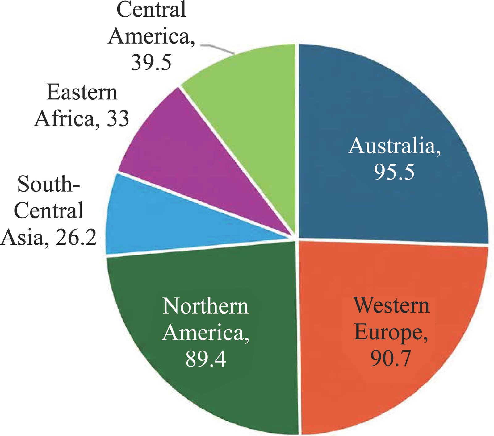
Fig. (1). Mortality rate of breast cancer in different countries.
The three predominant histological types that do occur sporadically are invasive lobular carcinoma (ILC), invasive ductal carcinoma (IDC), and mixed ductal-lobular carcinomas [3]. A kind of ductal carcinoma originates in the cells lining the conduits that transport milk from the glands to the nipple [1]. DCIS (ductal carcinoma in situ) exists in three grades, namely, low-grade (I), intermediate-grade (II), and high-grade (III), with a high probability of progressing to invasive ductal carcinoma [6]. DCIS, like IDC, has been subdivided into four intrinsic subtypes, including luminal A, luminal B, human epidermal growth factor receptor 2 (HER2/ERBB2)-positive, and basal-like breast cancer [7].
The HER2-positive subtype is found more frequently in DCIS than in IDC, present in around 35% of cases [6]. HER2-enriched subtypes overexpress as expected HER2-related genes and protein. Triple-negative breast cancer (TNBC), the most lethal and aggressive form of breast cancer, lacks ER, PR, and HER2 receptors [8]. The aggressive disease and poor prognosis of breast cancers are related to the abnormal over-expression of neu or c-erbB2/neu gene in breast cancers because of an increased transcription/translation of this transmembrane receptor tyrosine kinase [9].
The three most prominent techniques of dealing with breast cancer include chemotherapy (use of medicines to destroy cancer cells), radiotherapy (high-energy waves to destroy cancer cells), and surgery (the entire breast is removed or only the breast with cancer tissues is removed) [10–12]. Nevertheless, the same applies to radiation and chemotherapy, as they have some negative effects and do not yield better results when the time frame is longer [10]. To avoid adverse effects on healthy tissues and ensure effectiveness, several nanotechnological techniques are employed to deliver a potent anti-cancer drug selectively to breast cancer cells.
Through Nano carriers that are capable of navigating biological, biophysical, and biomedical barriers. Nano carriers probably provide a highly likely cure for breast cancer; it may be that small and versatile surface-modifying powers target areas of interest [12]. Most of the increasing number of nanoparticles (NPs) can be divided into either of two categories: 1. Organic cations consist of inorganic core ions, which are typically metals. 2. Organic molecules are the major component of organic anions [13]. Some of these organic compounds that it encapsulates include dendrimers, carbon nanotubes, emulsions, liposomes, and others. The basic structure that unites all the inorganic NPs consists of the electricity and the central core of the particle [14]. Forthcoming approaches in disease detection and management include the rapidly developing science of nanotechnology. Nanooncology is the subspeciality that concerns itself with the application in therapy and diagnosis [15]. Breast cancer metastasizes at a high rate to the lymph nodes and bones, which is why screening and diagnosis must be initiated at the earliest time possible [16]. The characteristics of nanoparticles employed for drug delivery systems are biocompatibility, self-assembly, stability, targeting, and encapsulation features [17]. Various NPs for tumor imaging and peripheral metastasis detection have been investigated with cancer-recognizing ligands attached to their surface [18].
Anthracyclines are effective in treating breast cancer at all stages, their use is restricted because large cumulative dosages of the drug carry a risk of heart damage [19]. When compared to unconjugated NPs and native rapamycin, EGFR-antibody conjugated NPs (EGFR-Rapa-NPs) demonstrated greater antiproliferative efficacy and higher cellular absorption in malignant breast cancer cells [20]. Comparably, the monoclonal antibody trastuzumab, which targets ERBB2 oncogene, improves the course of treatment for aggressive breast cancer but has a risk of cardiac damage, particularly in individuals who have received anthracycline therapy in the past [21]. Cancer cells can undergo apoptosis when Herceptin (trastuzumab) is coupled with quantum dots (QDs) for targeted bioimaging of HER2 receptors. Targeted anticancer therapies based on distinct tumor protein profiles can be achieved with silver or gold-containing and supermagnetic nanoparticles bioconjugates with antibodies against ERBB2 [22]. This article presents the targeted and new applicable methods for the treatment and control of breast cancer [12]. Many nanomaterials are undergoing clinical trials, and several of them have been successfully studied for their use in the treatment of cancer so far [23].
2. ER2-POSITIVE BREAST CANCER AND ITS PREVALENCE
HER2 positivity occurs in 20 to 30% of breast cancer cases and is due to the amplified HER2 gene that stimulates the growth of cancer cells through the overexpression of the HER2 protein [24]. HER receptors undergo dimerization and Tran’s phosphorylation, forming heterodimers with HER2 for activation, leading to cell angiogenesis, differentiation, proliferation, survival, and invasion. HER2-HER3 heterodimer stimulates PI3K/Akt, regulating cell growth and endurance [25]. HER2-positive customer malignancies typically provide an aggressive clinical picture and unsuitable prognosis; still, within ER and PR negative clients, being HER2 positive offers a superior prediction to Triple Negative Breast Cancer. The percentage prevalence of HER2-positive breast cancer in the pie chart represents that a major part of the world is suffering from HER2-negative breast cancer, which is almost 61% of the total, and the remaining part of the world is suffering from HER2-positive breast cancer, which is 26.20%, and low cancer, which is about 12-13%, respectively, as shown in Fig. (2) [26].
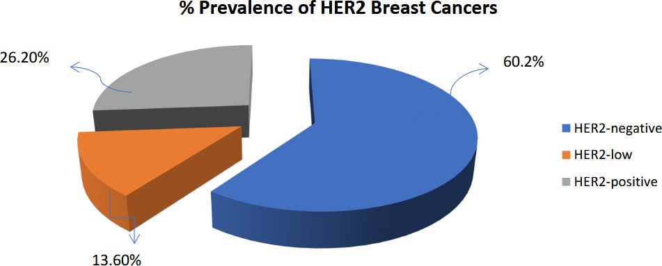
Fig. (2). The chart reveals the percentage prevalence of the patients: 60.2% were HER2-negative, 26.20% were HER2-positive, and 13.60% were HER2-low [26].
3. CONVENTIONAL DIAGNOSIS, TREATMENT, AND THEIR LIMITATIONS
Conventional diagnostic methods for HER2-positive breast cancer involve techniques, for instance immunohistochemistry (IHC), in-situ hybridization (ISH), Chromogenic in-situ hybridization, and Fluorescence in-situ hybridization (FISH), to spot HER2 protein. Specifically, immunohistochemistry applies a three-tier scoring system (from score zero to score 3+) to pinpoint the manifestation and strength of HER2 protein on the membrane [27]. Given the fact that HER2 amplification is directly linked to HER2 overexpression, in situ hybridization is applied when it is necessary to evaluate the number of copies of the HER2 gene in tumor nuclei [28]. Fluorescence in situ hybridization (FISH) is also used, which locates nucleic acids within cells using fluorescent probes [29]. Chromogenic in situ hybridization (CISH) is a cytogenetic method that uses a combination of in situ hybridization and a chromogenic signal detecting method of IHC [30]. CISH allows tissue slices to be preserved for extended periods without needing fluorescence microscopy, revealing considerable HER2 amplification [31]. Although these techniques are used for the diagnosis of HER2, unfortunately, there are some limitations, such as the fact that the results from fluorescence in situ hybridization. Tests can merely be viewed using a fluorescent microscope and cannot be stored to be analyzed later [32]. IHC scores of 0 and 1+ were deemed clinically insignificant, leading many pathologists to be unaware of their relevance, as shown in Table 1. Thus, the precise threshold definition of HER2 low positivity remains uncertain [33].
Table 1. Scoring of HER2 by IHC (immunohistochemistry) in accordance with (ASCO/CAP 2021) [7].
| Staining Pattern of Membrane | Tumor Cells | Scoring | Categories |
| Complete, Intense | >10% | 3+ | HER2+ |
| Complete, Weak-to-moderate | >10% | 2+ | HER2+ if ISH+ HER2- if ISH- |
| Incomplete, barely perceptible | >10% | 1+ | HER2- |
| Incomplete, barely perceptible | <10% | 0 | HER2- |
| No staining | – | 0 | HER2- |
Conventional treatment against HER2 involves the use of humanized monoclonal antibodies against HER2, which have significantly enhanced the management of HER2-positive breast cancer by downsizing tumors, thus improving receptivity [34]. Before surgery, customized options are offered by dual anti-HER2 therapy and chemotherapy, depending on the specifics of the tumor and the response of the individual patient. HER2-targeted therapies have proven effective against HER2-positive breast cancer, but resistance often emerges through complex molecular pathways, weakening treatment effectiveness and allowing illness recurrence or tumor growth to continue unchecked, as visible in Fig. (3) [35]. Intracellular sources of acquired drug resistance in breast cancer are mutations in P53, overexpression of ABC transporters, amplification of HER2, upregulation of permeability glycoprotein (P-gp), enhanced activity of thymidylate synthases, and aberrations in BRAC1 [37].
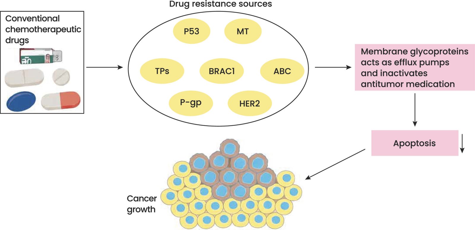
Fig. (3). The relationship between drug resistance sources and tumor cells for conventional chemotherapy medicines [37] (reproduced with permission).
4. TARGETED DELIVERY OF ANTI-HER2 DRUGS AND LIGANDS
Cancer therapy is challenged by the drug delivery of chemotherapy medication, contrast agents, and radionuclides to the tumor site. Targeted therapy offers cancer treatment through a targeted approach without involving healthy cells. Reversible dual tyrosine kinase inhibitor lapatinib is an inhibitor that binds and blocks epidermal growth factor receptor (EGFRs) activity and HER2 receptor [38].
4.1. HER2-Specific Ligands for Targeted Drug Delivery
Trastuzumab, a humanized recombinant monoclonal antibody, is directed against HER2 and used for the treatment of HER2+ breast cancer [39]. The effectiveness of this monoclonal antibody depends on HER2 expression on the tumor, mutation, and eventual resistance. Some known mechanisms include mutation of PI3K, increasing activity of other TK receptors, loss of the extracellular domain of HER2, and inactivation of the endocytic adapter protein endophilin A2, leading to inefficient internalization of HER2 [40]. One of the inhibitors, lapatinib, is a reversible dual tyrosine kinase inhibitor that interacts and inhibits epidermal growth factor receptor (EGFR) or HER2 receptor [41]. Pertuzumab is a humanized monoclonal antibody targeting HER2 on the extracellular domain II, inhibiting the formation of homo and heterodimers, and preventing HER2/HER3 kinases[42]. Co-administration of Pertuzumab with trastuzumab is observed to be more operative for HER2+ breast cancer, and the recommendation for first-line treatment of HER2+ BC is the grouping of two monoclonal antibodies and taxanes [43]. However, resistance to pertuzumab is related to S310F mutations, EGFR-HER3 heterodimers, and regulation by miRNA [44]. These drugs are designed together with a special peptide that binds to the tumor in order to lessen the number of side effects and achieve the optimal concentration in the sick area [45]. Four peptide sequences, P51, P25, P47, and P40, can be used for developing imaging agents/or targeted drug delivery in HER2-positive breast cancer [46].
4.2. Antibody Drug Conjugates ADCs
ADCs, a type of protein-based natural medicinal product to control cancer, are composed of anticancer potential assigned with the stability, target-selecting tendency, and strength of monoclonal antibodies (mAbs) [40, 47, 48]. An ADC conjugated to the humanized antibody trastuzumab is known as trastuzumab emtansine (T-DM1). It contains the maytansinoid toxin DM1 [49]. It minimizes side effects to other tissues because it only releases its drug through the receptor-mediated endocytosis mechanism [51]. The possible mechanisms of T-DM1 resistance are the downregulation of HER2, alteration in the internalization of the HER2-T-DM1 complex, and the inhibited lysosomal release of lysine-MCC-DM1 [52]. ADC trastuzumab deruxtecan effectively cleaves lysosomal cathepsin to release cytotoxic medications [42, 53]. T-DXd has good ADME properties, and its drug-to-antibody ratio is higher. Payload mechanisms and monoclonal mechanisms are some of the strategies of resistance. The overall survival and the progression-free survival rates are positively changing after the T-DM1 treatment and are sensitive to the ERBB2 mRNA expression. The DAISY study also found that recurrent mutations inside the SLX4 gene may affect T-DXd resistance [40, 53].
TYPES OF NANOPARTICLES USED IN THE DIAGNOSIS AND TREATMENT OF HER2-POSITIVE BREAST CANCER
5.1. Gold Nanoparticles (GNPs)
Gold Nanoparticles (GNPs) can be applied to cancer radiotherapy for the destruction of cancerous cells by binding them to monoclonal antibodies (mAbs). To efficiently detect the tumor using a single specific mAb, HER2, a protein engineering technique was applied to conjugate permeation, to the C-terminus of a broad-type anti-HER2 antibody heavy chain, a cell-penetrating peptide (CPP) [56]. One mAb-2CPP maintained the capability to bind HER2 and efficiently mediate the internalizing capability of the antibody in MCF-7 breast cancer. The MAb-2CPP-GNP complex produced a higher death rate than mAb-GNP [57]. A novel multimodal radio-bioconjugate containing doxorubicin (DOX) α-emitter (198Au)-based targeted therapy of cancer tissues with HER2-positive receptors has been developed utilizing trastuzumab as a guiding vehicle. The produced nanoparticles that contained a poly (ethylene glycol) linker & a high affinity to bind to HER2 receptors were absorbed by cells [59].
5.2. Quantum Dots
Carbon quantum dots (CQDs) enhanced with nitrogen were employed in an immunoelectrode to detect the breast cancer biomarker, HER2 [60–62]. Quantitative characterization was noticed as more effective in the quantum dot-based immunofluorescence approach compared to the usual diagnostic methods [63]. For the voltametric study of breast cancer cells and HER2-ECD in human serum, an assay of immunomagnetic beads conjugated with streptavidin and CdSe@ZnS quantum dots was designed. They noted that this method presents a sound paradigm in the identification of breast cancer [60]. A novel method of staining to herd quantitative data on HER2 in breast cancer tissues is quantum dot extending trastuzumab immunohistochemistry [66]. By linking fluorescence levels to quantum dot concentrations, this approach advances cancer detection and therapy planning [61]. The CdSe/CdZnS quantum dots have emerged as useful sensors for reliably targeting cancer biomarkers [64, 65]. When conjugated to hyaluronic acid, N-GQDs present minimal toxicity and enhanced fluorescence for the detection of cancer cells. While sensitive, CdSe/ZnS quantum dots need to be validated and tested for approximating quantification of their time-dependent use in therapy effects [66]. Although PEGylated QDs offer enhanced photostability and imaging severity, the binding efficiency may be altered due to the presence of a polyethylene glycol moiety [67]. Uses of QDs can be quantified to determine levels of HER2 expression in cells, improve the sensitivity and staining of detection, and help with the quantitative characterization of breast cancer subtypes. While the tagging efficiency of these dots is very high (83%), the impact of these dots on cell survival remains rather obscure. To obtain a better signal for the examined results, solid-state zinc-adsorbed carbon quantum dots immobilized on gold nanoparticles, as well as magnetic beads, are employed [61]. The graphite sheet substrate was used to fabricate the immunoelectrode that helps to sense and assess the HER2 biomarker on breast cancer with nitrogen-coated carbon quantum dots (N-CQDs). The bovine serum albumin (BSA)-modified immunoelectrode immunodefinitive method has the capability to help in detecting HER2 at high sensitivity and specificity rate and therefore results in highly accurate HER2 diagnosis in blood samples (Fig. 4) [61]. A thorough review of the existing literature shows a significant gap between diagnostic sensitivity and therapeutic feasibility of quantum dots. Although the platforms based on N-CQDs show excellent detection limits at picogram level in human serum, the persistence of heavy-metal cores in the system over time is a serious obstacle to the implementation in live therapeutics. Future studies should focus more on the long term clearance rather than analytical validation.
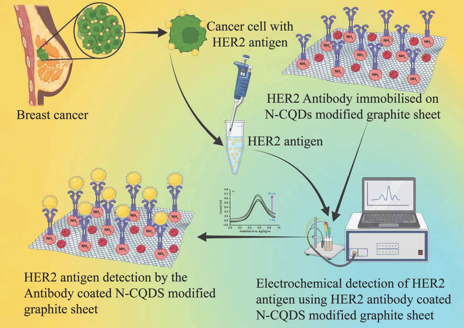
Fig. (4). Shows a diagram of nitrogen-functionalized carbon quantum dots (N-CQDs) on a sample of coated graphite sheet (GS) in relation to electrochemical detection of breast cancer biomarker: Human epidermal growth factor receptor2 (HER2). A hyperstable bovine serum albumin version of this N-CQDS/GS -modified HER2 antibody specific to untreated blood samples, the addition has a linear response range and a low detection limit of 0.1-1ng/mL and 4.8pg/mL, respectively [61] (reproduced with permission).
5.3. Immunoliposomes
It was said that liposomal formulations for the treatment of breast tumors have merits, while they have demerits such as short circulation time in vivo and low bioavailability due to low concentration within the target malignant cells [68]. PEGylated liposomes are formed through the conjugation with polymers like polyethylene glycol and exhibit enhanced in vivo circulation times and better tumor tissue uptake [69]. In recent one-decade, a considerable number of publications have focused on the use of specialized immunoliposomes for the purpose of treating breast cancer due to the overexpressed cell surface receptors such as HER2 [70, 71]. Liposomal formulations containing rapamycin and immuno-liposomes containing trastuzumab for improving the therapeutic outcome in HER2+ breast cancer [72–74]. Targeting of rapamycin to the trastuzumab-coated surface was done through immunoliposomes which showed high efficiency and stability in antibody functionalization [75].
Paclitaxel (PTX) conjugated to trastuzumab & Elacridar (ELA)-peptidoglycan PEGylated pH-sensitive liposomes (TPPLs) as targeted anticancer drug carriers [76, 77]. PTX is used as an anticancer drug, but single use contributes to MDR and therapy failure. They administered combined PTX with their selective P-glycoprotein inhibitor, ELA, so that they could have maximal therapeutic effects [78]. In vitro drug release studies indicated that it is possible to obtain a better percentage of drug release at a pH of 5 rather than a pH of 7.4, which indicates the physiological pH. In vivo tumor regression studies demonstrated that TPPLs reduced a greater amount of tumor burden, had lower toxicity levels, and were more effective against tumors than their corresponding PPLs [79]. In tumor models, pH-sensitive TPPL immunoliposomes exhibit improved regression dynamics, but one of the key challenges is the degradation of the lipid bilayers during the scale-up manufacturing process. The clinical failure of many liposomal formulations is not due to poor targeting efficacy, but rather to variations in the number of ligands attached to each formulation, as well as to variability in the amount of ligands encapsulated within each formulation.
5.4. Silica-Based Nanoparticles (SiNPs)
The delivery of chemotherapy drugs and in vivo imaging can be done at the same time, without invasiveness, when using Silica-based nanoparticles (SiNPs) for HER2-positive breast cancer treatment [80, 81]. The modified SiNPs were able to load due to Doxorubicin. SiNPs of two ratios of Trastuzumab half-chain, Hc-TZ to one nanoparticle (approximately) are harvested and used as the linker. These SiNPs were radiolabeled at histidine residues with 99mTc [21]. There was a reduction of tumor volume and mass at the end of therapy; the nanoparticles showed comparable DOX carrying abilities as liposomal doxorubicin (Caelyx) [81, 82]. Fig. (5) depicts that SiNPs-TZ were stained with the FITC dye to distinguish the nanoparticles in vivo. The synthesis and characterisation of Hc-TZ functionalised SiNPs-TZ was performed by a 1:8 ratio of SiNPs to Hc-TZ. Reacting protocols were done through a fifth of the Hc-TZ dose to get this figure to two Hc-TZ per nanoparticle [81].
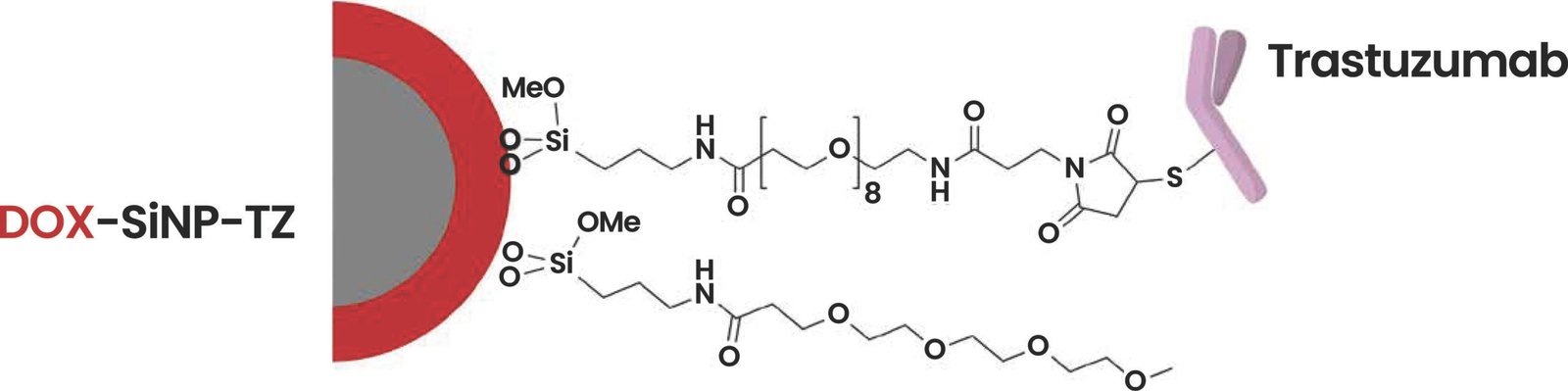
Fig. (5). Targeted (Hc-TZ) spherical silica nanoparticles DOX-SiNPs-TZ (hydrodynamic diameter ~70-80nm). The nanoparticles’ silica core (grey) was highly loaded with doxorubicin (red) [81] (reproduced with permission).
These nanomedicines are not to be considered as single drugs, but rather a comparative assessment is needed to understand which chemical structures will be best suited for clinical scale-up, which is critically analyzed in Table 2.
Table 2. Comparative analysis of nanocarrier modalities in HER2-positive breast cancer therapeutics.
| Nanoparticle Type | Key Mechanism of Action | Practical Advantages | Critical Limitations & Bottlenecks |
| Gold Nanoparticles (GNPs) | Radiosensitization; heavy-chain cell-penetrating peptide antibody conjugation. | High surface-to-volume ratio; excellent photothermal conversion. | Non-biodegradable; long-term hepatic and splenic accumulation toxicity. |
| Quantum Dots (QDs) | Immunofluorescence; electrochemical biosensing via functionalized graphite sheets. | High quantum yield; extreme sensitivity for low-limit antigen detection. | Potential cytotoxicity from heavy metal cores (e.g., CdSe); photoblueshift risks. |
| Immunoliposomes | Receptor-mediated endocytosis of encapsulated payloads (e.g., PTX, Rapamycin). | Highly biocompatible; prolonged in vivo circulation when PEGylated. | Low structural stability; risk of premature drug leakage before target localization. |
| Silica-based Nanoparticles (SiNPs) | Simultaneous therapeutic delivery (DOX) and 99mTc-radiolabeled in vivo diagnostic imaging. | Rigid, stable pore architecture; high payload capacity; ideal for theranostics. | Slow degradation kinetics; potential for immune clearance via the mononuclear phagocyte system. |
6. DIAGNOSIS OF HER2-POSITIVE BREAST CANCER USING NANOTECHNOLOGICAL STRATEGIES
Current diagnostic methods, which determine treatment approaches for HER2 levels, have flaws. [83, 84]. Accurately gauging patients’ HER2 is pivotal so physicians can craft suitable remedies, including immunohistochemistry, chromogenic in situ hybridization, and fluorescence in situ hybridization normally used [85]. However, these procedures aren’t flawless [86]. Around 20% of HER2 classification by standard means are later found to be inaccurate [87, 88]. Novel techniques targeting HER2 might aid earlier disease identification, guide options for care, and extend survival rates. Recently, organic and inorganic nanoparticles, quantum dots, and aptamers in particular have shown promise. Innovative imaging employing HER2-targeted nanoparticles allows oncologists to properly judge its presence and develop the right strategies compared to traditional means [89]. Imaging modalities have been applied as positron emission tomography, computerized tomography, optical imaging, single photon emission computed tomography, magnetic resonance imaging, and ultrasounds, as shown in Fig. (6) [90, 91]. Positron emission tomography and single photon emission computed tomography are more sensitive than magnetic resonance imaging, but offer poorer resolution and lack an anatomical framework [92].
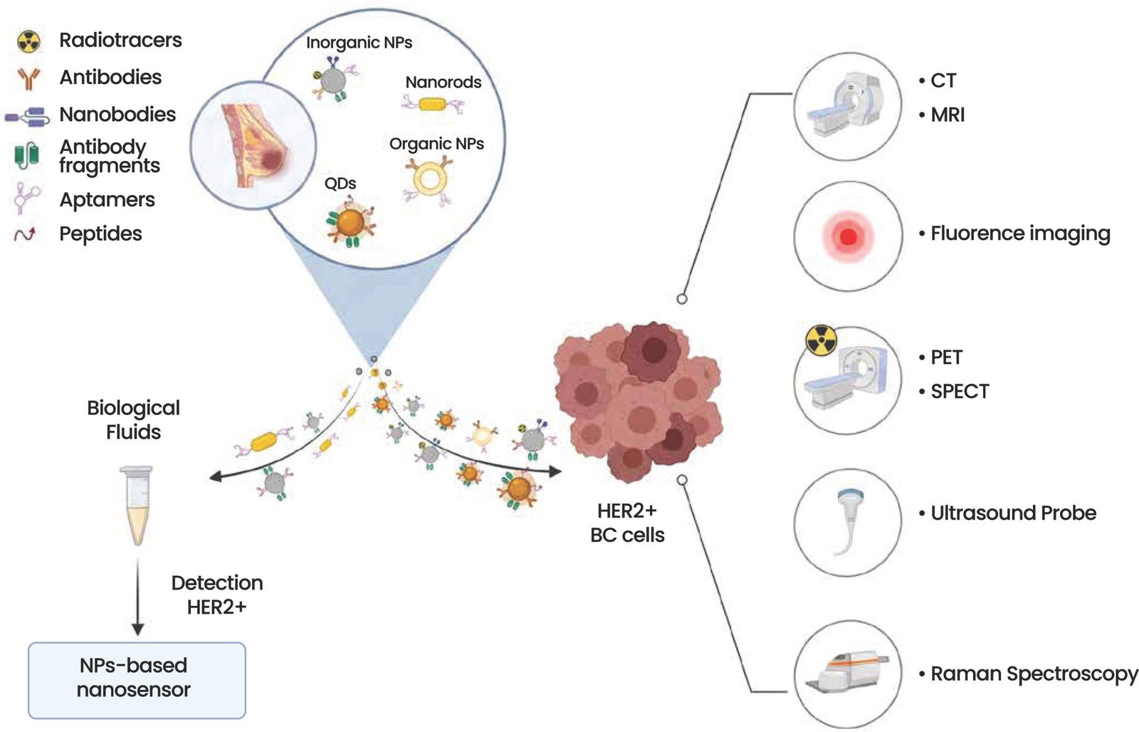
Fig. (6). Approaches of nanotechnology aimed at the diagnosis of her2 positive breast cancer [114] (reproduced with permission).
6.1. Nuclear Magnetic Resonance and Computer-Aided Tomography
Magnetic resonance imaging (MRI) is an invasive imaging technique for diagnosing tumors because it has high soft tissue contrast and high spatial resolution, in contrast to signal depth, with the use of exogenous contrast mediums injected before the scan. This study envisaged using dextran-modified superparamagnetic iron oxide nanoparticles conjugated with Herceptin for the recognition of HER2 both on a tissue microarray and in a tissue culture of MCF-7 cells [93]. The authors described marked improvements in magnetic resonance for numerous BC cell types, and in vivo, the contrast agent deposition was verified for the recognition of HER2 breast tumor [94]. PEGylated SPIONs, single chain variable fragment, and a fluorescent tag of Cyanine-based to functionalize particles for dual targeting were used to target HER2-Positive breast cancer cells, with increased uptake proportional to the level of HER2 expression [95]. At present, these particles, due to their biocompatibility and biodegradable nature are suitable for the uncovering of the early stage of breast cancer.
6.2. Multimodal Hybrid Methods
The capability of combining multiple imaging methods on a nano-scale platform, employing silica-coated magnetic nanoparticles that enabled both fluorescence microscopy and magnetic resonance imaging in vitro, was explored [22]. The particles, functionalized with trastuzumab, provided a striking contrast when detecting HER2 expression on SKBR-3 cells, indicating potential as agents for further multimodal in vivo examination. Additionally, iron oxide nanoparticles with fluorescent labels and an engineered antibody for targeting HER2 receptors on cells and tumors are under laboratory conditions and within living subjects [96]. They found the customized nanoparticles intensely bound to SKBR-3 breast cancer cells overexpressing HER2 but barely attached to MDA-MB-231 cells deficient in the receptor, as observed in mouse models [97]. Moreover, iron oxide nanoprobes carrying a cyanine dye, polyethylene glycol polymers, and an anti-HER2 single-chain antibody fragment showed a marked amplification of magnetic resonance signals in mice bearing BT474 tumors 24 hours post-injection, demonstrating specificity for cancer cells with high HER2 levels over unaffected cells [95].
6.3. Inorganic NPs Based Nano Sensors
Nano-biosensors are used to govern the concentration of HER2 in patient blood and tissue specimens to diagnose early HER2-Positive BC, predict recurrence, metastasis, and enhance treatment regimen in low response or resistant cases [98]. One of the scientist Villegas-Serralta et al., synthesized amino silane covalently functionalized and dextran surface-covered magnetic nanoparticles (As-M and Dx-M) coupled by a Single-Chain Fragment Variable (scFv) of anti-HER2 monoclonal antibody to determine their feasibility as magnetic nano-biosensors used in HER2 detection employing a magnetic enzyme-linked immunosorbent assay (m-ELISA) [99]. Raman spectroscopy could also detect As-M, which is a helpful method in HER2 quantification and recognition [100]. Nano Based platform was synthesized through gold nanoclusters encapsulated in liver oil, and have been conjugated with an Aptamer or anti-HER2 antibody [101]. The scientists verified that their comparatively cheap Nano-biosensor is virtually non-toxic to the cells, effectively measures the concentration of HER2 in cell emulsions, as well as individualizes cancer cells with HER2 receptor on the section of tissue [102].
6.4. Aptamers-Based Nano Sensors
Aptamer-based organic nanoparticles display promising potential for identifying HER2 positive tumor cells in tissues and fluids. In the studies, two Aptamer of DNA named HeA2_3 and HeA2_1 performed the whole-cell SELEX (systematic evolution of ligands by exponential enrichment), which possess considerable specificity to bind with HER2; they also investigated that these aptamers bind highly towards HER2-Positive cell line and tumor tissues in contrast to the cell line MDA-MB-231 with comparatively low expression of HER2 [103]. Additionally, Kim and co-workers evolved aptamers for plasmonic nano sensors intended for detecting HER2 in biological fluids. They affixed anti-HER2 positive targeting Aptamers to gold Nano rods, showcasing a less limit of recognition and paving the means for highly delicate diagnostic techniques [104].
7. NANOTECHNOLOGICAL STRATEGIES FOR THE TREATMENT OF HER2-POSITIVE BREAST CANCER
7.1. Approaches to the Administration of Chemotherapeutic Agents and Other Cytotoxic Compounds
Lately, several types of NPs functionalized with targeting ligands that recognize HER2 have been employed for various drugs in HER2+ BC. Non-targeted docetaxel-loaded TZ-coated polymer-based NPs remarkably enhanced the target selectivity, cellular adhesion, and cytotoxic effect on HER2-Positive BT474 cells but not on the HER2 control cells [105]. Although in general polymer-based NPs and cisplatin-loaded lipid had a high in vitro efficiency in contrast to free drug and non-targeted NPs, it was specific for HER2-Positive SKOV-3 but not for HER2-Negative HCC70 cells [106]. Doxorubicin (DOX), widely used for the treatment of HER2-Positive BC, has been incorporated into the antibody functionalized polymeric NPs and demonstrates to be more cytotoxic to HER2-Positive MCF7 cancer cells than DOX-NPs without antibody and free DOX, but without harming normal cells [107]. In a parallel therapeutic approach designed to bypass conventional multi-drug resistance, PLGA (Polylactic-co-glycolic acid) NPs coupled with HER2 nano body and encapsulating saporin targeted specifically to SKBR-3 cells. This method employs a local light exposure and photosensitizer to stimulate the release of the encapsulated nanocarriers at the endosomal level [108]. Nanoformulation of Curcumin, synthesis, and application for BC management through SKBR-3 human serum albumin with NP adorned by a link to anti HER2 aptamer [109].
7.2. Approaches to Nucleic Acid Conveyances or Gene Silencing Molecules
Small interfering RNA (siRNA) is a type of double-stranded RNA non-coding molecule. When using siRNAs, it becomes easier to knock down certain genes and treat cancer of certain targets but they cannot be drugged. Nevertheless, it became clear that siRNAs cannot be used in vivo as such because of their inherent instability [110]. To address resistance issues, mesoporous silica nanoparticles were designed that were functionalized with conjugate HER2 targeting antibodies to carry HER2 siRNA; it was found that HER2-Positive BC cells very rarely acquire fighting to HER2 siRNA as opposed to Lapatinib or TZ. The siRNA could really turn off the expression of HER2 at mRNA level, and this cannot be offset by any adaptive changes and survival mechanisms [110]. In a study, a mini Nano drug based on polymeric acid was applied as a distribution system for peptides and antisense oligonucleotides HER2-specific that target the BT474 cells expressing HER2 receptors [38].
7.3. HER2-Positive Tumors: Nanoparticle-Based Strategies to Enhance Irradiation Efficiencies
Radiotherapy in the form of α-Nano brachytherapy is employed in early and advanced stages of breast cancer, along with the management of metastatic cancer-related pain. This approach involves introducing Au nanoparticles conjugated with 211Atemitter and illuminating the bismuth substrates. The NPs are kept stable using PEG and functionalized with Tinidazole (TZ) for targeting purposes. In vitro, the evidence shows that 211At-AuNPs-PEG-TZ has potential in the treatment of HER2+ cancers, as evidenced by experiments conducted on SKOV-3 ovarian cancer cells [21]. Partial thromboplastin time (PTT) is a non-surgical treatment wherein photo-thermal agents are applied to reduce the dimensions of the tumor mass. Conjugated NPs with enhanced affinities for tumors and high PTT conversion efficiency are considered more effective for cancer treatment. Gold Nano rods, whose size and shape can be controlled to achieve desired near infrared wavelengths of absorption, display certain properties, making them ideal photo thermal agents [111]. However, the Nano rods made up of plain gold cannot selectively mark the lump, and consequently, their ability to treat is hindered. Consequently, special gold Nano rods (GNRs) operationalized with porphyrin and TZ were developed; the enzymes show a higher targeting specificity and stronger anti-tumor effect in a BT474 heterograft in vivo model [112]. The study showed clearly that GNRs have potential application for HER2+ BC and DARPins (designed ankyrin repeat proteins) can enhance the UCNPs (upconverting nanoparticles) uptake by SKBR-3 HER2+ cells for PTT mediators. This method was also verified in vivo in the lung cancer mouse model of Lewis, where functionalized UPNCs provided the shrinkage of the tumor size after a single laser irradiation [113].
CLINICAL TRIALS
The clinical management of HER2-positive breast cancer is heavily challenged by profound intratumor and intertumor heterogeneity [114]. Considering that metastatic HER2-positive breast cancer remains fatal, it is still a key clinical problem at this time. New products targeting HER2 have enhanced early and later stage general survival considerably. However, de novo or acquired resistance enables a metastatic HER2-positive breast cancer patient to take anticipatory action in due course [115]. Around 20% of the breast cancer patients are HER2 positive, while others are HER2 negative, and they range from 70-80%. Furthermore, in the early stage, it is assessed that about 5-10% of the affected role diagnosed with breast cancer demonstrate distant metastases at the time of diagnosis. CDK4/6 inhibitors significantly improve prognoses, making them standard of care alongside endocrine therapy for these patients [116]. The Ministry of Health drug program in Poland only allows co-financing of CDK4/6 inhibitors for patients, potentially delaying their use or relying on cheaper, less efficient drugs from older generations [117].
FUTURE DIRECTIONS
Although breast cancer is the second leading cause of death among women, there has been a decrease in deaths due to early diagnosis instruments and new methods of treatment. Breast cancer contributed to 24.5 percent of the new cases and 15.5 percent of deaths due to cancer in 2020 only [19]. More formulations are still coming to clinical trials, and in the near future, other approvals are expected. It is noted that in 15-20% of cases with invasive breast tumors, the HER2, or ERBB2 gene, is amplified or upregulated. This has been linked to an aggressive phenotype and, accordingly, poor prognosis [118]. Bench-to-bedside translation of oncology involves initiation of HER2-targeted therapies like tyrosine-kinase inhibitors, monoclonal antibodies, and antibody-drug conjugates [119]. But there are still several challenges that need to be overcome, among these are the toxicities of nanoparticles, uncontrolled release of drugs, and the potential of immune cells to clear nanoparticles [120]. The conceptual model identifies factors contributing to resistance to HER2 treatments in tumors overexpressing HER2, including intratumoral heterogeneity, immune evasion, and potential future approaches for improved patient outcomes [121]. Although breast cancer is still one of the main cancers worldwide, the death rate from breast cancer is slowly decreasing with the advancement of diagnosis and targeted front-line therapies [122]. But, the clinical translation of advanced nanomedicines is hindered by nanoparticle toxicity, fast clearance by the immune system, and scalability bottlenecks in cGMP [123]. Smart, stimuli-responsive platforms co-delivering anti-MDR microRNAs and chemotherapeutics are key to future success, which will be able to programatically overcome efflux pumps and tumor heterogeneity [124].
CONCLUSION
The application of nanotechnology in the management of HER2-positive breast cancer represents a paradigm change from non-specific and highly toxic conventional therapies to molecular targeted therapy. Successful enhancement of the therapeutic index of potent chemotherapeutic agents, gene-silencing molecules, and contrast media can be achieved by engineering of organic and inorganic nanocarriers with anti-HER2 antibodies, peptides or aptamers. This specific design reduces the amount of off-target cardiotoxicity, a major problem that has been a concern in the past with free anthracyclines and trastuzumab treatments. Yet, nanomedicine is not a panacea; for it to be successful in the clinic, some critical translational challenges must be overcome, such as the biophysical behavior of the protein corona, scale-up manufacturing consistency, and long-term tissue bioaccumulation toxicity. Future clinical benchmarks will be based on multi-targeted, stimuli-responsive systems that will be able to dynamically respond to the tumor microenvironment. The continued interdisciplinary cooperation of molecular oncologists, materials scientists, and clinical trialists is a must to make these sophisticated lab prototypes into life-saving bedside realities.
LIST OF ABBREVIATIONS
BSA | = | Bovine Serum Albumin |
CISH | = | Chromogenic In Situ Hybridization |
CPP | = | Cell-Penetrating Peptide |
CQDs | = | Carbon Quantum Dots |
DARPins | = | Designed Ankyrin Repeat Proteins |
DCIS | = | Ductal Carcinoma in Situ |
DOX | = | Doxorubicin |
EGFR-Rapa-NPs | = | EGFR-Antibody-Conjugated NPs |
EGFRs | = | Epidermal Growth Factor Receptors |
FISH | = | Fluorescence In Situ Hybridization |
GNPs | = | Gold Nanoparticles |
GNRs | = | Gold Nano rods |
GS | = | Coated Graphite Sheet |
HER2 | = | Human Epidermal Growth Factor Receptor 2 |
IHC | = | Immunohistochemistry |
IDC | = | Invasive Ductal Carcinoma |
ILC | = | Invasive Lobular Carcinoma |
m-ELISA | = | Magnetic Enzyme-Linked Immunosorbent Assay |
mAbs | = | Monoclonal Antibodies |
MRI | = | Magnetic Resonance Imaging |
N-CQDs | = | Nitrogen-Coated Carbon Quantum Dots |
N-CQDs | = | Nitrogen-Functionalized Carbon Quantum Dots |
NPs | = | Nanoparticles |
P-gp | = | Permeability Glycoprotein |
PLGA | = | Polylactic-co-Glycolic Acid |
PTT | = | Partial Thromboplastin Time |
PTX | = | Paclitaxel |
QDs | = | Quantum Dots |
scFv | = | Single Chain Variable Fragment |
SELEX | = | Systematic Evolution of Ligands by Exponential Enrichment |
SiNPs | = | Silica-Based Nanoparticles |
siRNA | = | Small Interfering RNA |
T-DM1 | = | Trastuzumab Emtansine |
TNBC | = | Triple-Negative Breast Cancer |
TZ | = | Tinidazole |
UCNPs | = | Upconverting Nanoparticles |
AUTHORS’ CONTRIBUTION
SHH: Supervision, Investigation Author.
MH: Writing – review & editing; Lead; Formal Analysis.
AA: Validation; Formal analysis; Writing – review & editing: Equal; Writing – original draft.
SK: Writing – review & editing: Equal; Visualization.
AY: Data curation; Methodology.
NL: Writing – review & editing: Conceptualization.
KI: Data curation; Visualization.
MS: Methodology; Writing – review & editing.
TJ: Formal Analysis.
HI: Supervision.
CONSENT FOR PUBLICATION
Not applicable.
AVAILABILITY OF DATA AND MATERIALS
Not applicable.
FUNDING
This research did not receive any specific allowance from funding agencies in the public, commercial, or not-for-profit sectors.
CONFLICT OF INTEREST
The authors declare that they have no competing interests for the publication of this review article.
ACKNOWLEDGEMENTS
We would like to express our deepest gratitude to the Department of Biochemistry & Biotechnology at the University of Gujrat for their significant contributions and support for this project. We also extend our thanks to our mentors for their invaluable guidance and to our colleagues for their encouragement throughout this work.
DECLARATION OF AI
During the preparation of this work the authors used ChatGPT for editing purposes. After using this tool, the authors reviewed and edited the content as needed and take full responsibility for the content of the published article
REFERENCES
[1] Obeagu EI, Obeagu GU. Breast cancer: A review of risk factors and diagnosis. Medicine. 2024;103(3):e36905.
https://doi.org/10.1097/MD.0000000000036905
[2] Grattagliano Z, Grattagliano A. Breast Cancer: The Road to a Personalized Prevention. Igmin Res. 2024;2(3):163-70.
https://dx.doi.org/10.61927/igmin160
[3] Tseu GYW, Kamaruzaman KA. A review of different types of liposomes and their advancements as a form of gene therapy treatment for breast cancer. Molecules. 2023;28(3):1498.
https://doi.org/10.3390/molecules28031498
[4] Davis SC, Snyder E. Factors impacting quality of life for breast cancer survivors. Nurse Pract. 2024;49(5):17-23.
https://doi.org/10.1097/01.NPR.0000000000000172
[5] Xu Y, Gong M, Wang Y, Yang Y, Liu S, Zeng Q. Global trends and forecasts of breast cancer incidence and deaths. Sci Data. 2023;10(1):334.
https://doi.org/10.1038/s41597-023-02253-5
[6] Wang J, Li B, Luo M, Huang J, Zhang K, Zheng S, et al. Progression from ductal carcinoma in situ to invasive breast cancer: molecular features and clinical significance. Signal Transduct Target Ther. 2024;9(1):83.
https://doi.org/10.1038/s41392-024-01779-3
[7] Theivendren P, Pavadai P, Kunjiappan S, Ravi K, Kiruthiga N, Chidamabaram K, et al. Emerging therapeutic strategies and opportunities in targeting protein pathways for breast cancer treatment: a critical review. Nanotechnology. 2025.
https://doi.org/10.1088/1361-6528/add6ae
[8] Mir MA, Qayoom H. Introduction to breast cancer. In: Therapeutic potential of cell cycle kinases in breast cancer. Singapore: Springer; 2023. p. 1-22.
https://doi.org/10.1007/978-981-19-8911-7_1
[9] Prat A, Pascual T, De Angelis C, Gutierrez C, Llombart-Cussac A, Wang T, et al. HER2-enriched subtype and ERBB2 expression in HER2-positive breast cancer treated with dual HER2 blockade. J Natl Cancer Inst. 2020;112(1):46-54.
https://doi.org/10.1093/jnci/djz042
[10] Arshad R, Kiani MH, Rahdar A, Sargazi S, Barani M, Shojaei S, et al. Nano-based theranostic platforms for breast cancer: a review of latest advancements. Bioengineering. 2022;9(7):320.
https://doi.org/10.3390/bioengineering9070320
[11] Tran P, Lee SE, Kim DH, Pyo YC, Park JS. Recent advances of nanotechnology for the delivery of anticancer drugs for breast cancer treatment. J Pharm Investig. 2020;50:261-70.
https://doi.org/10.1007/s40005-019-00459-7
[12] Addissouky T, El Tantawy El Sayed I, Ali M, Alubiady M, Wang Y. Harnessing innovation for the future of breast cancer management. Clin Res Oncol. 2024;1(1):10-7.
https://doi.org/10.46439/oncology.1.004
[13] Bhandari M, Raj S, Alam MS. Recent innovations in nanomedicine and nano-based techniques for the treatment of breast cancer. BioImpacts. 2025;15:30804.
https://doi.org/10.34172/bi.30804
[14] Fernandes CL, Silva DJ, Mesquita A. Novel HER-2 targeted therapies in breast cancer. Cancers. 2023;16(1):87.
https://doi.org/10.3390/cancers16010087
[15] Aljadani M. Nanotechnology-based therapeutic strategies for breast cancer. Int J Pharm Investig. 2022;12(2).
https://doi.org/10.5530/ijpi.2022.2.27
[16] Colombo M, Corsi F, Foschi D, Mazzantini E, Mazzucchelli S, Morasso C, et al. HER2 targeting as a two-sided strategy for breast cancer diagnosis and treatment: Outlook and recent implications in nanomedical approaches. Pharmacol Res. 2010;62(2):150-65.
https://doi.org/10.1016/j.phrs.2010.01.013
[17] Wang T, Cao C, Gavahi M, Sadri M, Rong X. Using nanotechnology for diagnosis and treatment of breast cancer: A review. Indian J Pharm Sci. 2022;84.
https://doi.org/10.36468/pharmaceutical-sciences.spl.480
[18] Fakudze N, Chota A, Abrahamse H, George BP. Nano-theragnostic platforms for breast cancer diagnosis and therapy: Current advances, translational challenges, and future directions. In: Nano theragnostics in breast cancer: Advances, challenges, and future prospects. Singapore: Springer; 2026. p. 789-815.
https://doi.org/10.1007/978-981-95-3682-5_21
[19] Chattopadhyay A, Goyal F, Sehrawat A, Sidhu IS, Monga V, Bhatti GK, et al. Revolutionizing breast cancer therapeutics: Intersecting frontiers of precision medicine, nanotechnology, and drug delivery innovations. Curr Treat Options Oncol. 2025;26(9):775-96.
https://doi.org/10.1007/s11864-025-01343-3
[20] Talaee O, Mehrabifard M, Alipour B, Derakhshan S, Asghariazar V, Zamani H, et al. Characterization of a HER-2 targeted silica-gold nanoshells for breast cancer radio-photothermal therapy. Appl Radiat Isot. 2026;112454.
https://doi.org/10.1016/j.apradiso.2026.112454
[21] Wu W, He Y, Zhang C. Emerging Targeted and Multimodal Therapeutic Strategies in Breast Cancer: A Comprehensive Review. Breast Cancer Targets Ther. 2026;18:1-28. https://doi.org/10.2147/BCTT.S575936
[22] Sonalisha S, Kirti A, Asima S, Parashar R, Pal S, Barik D, et al. Translational paradigm of Advanced Nanoscale Strategies for Triple Negative Breast Cancer (TNBC): Mechanistic Insights, Metastatic Pathways, and emerging theragnosis. Mater Adv. 2026. https://doi.org/10.1039/d5ma00866b
[23] Kim A, Mo K, Kwon H, Choe S, Park M, Kwak W, et al. Epigenetic regulation in breast cancer: Insights on epidrugs. Epigenomes. 2023;7(1):6.
https://doi.org/10.3390/epigenomes7010006
[24] Rojo F, Calvo-Martinez L, Bernet L, Burgués O, García-Caballero T, Cobo SLT, et al. 281P Prevalence of HER2-low breast cancer in the GEICAM/2011-06 trial: Agreement in HER2-low classification between standardized immunohistochemistry assays. Ann Oncol. 2024;35:S332-3.
https://doi.org/10.1016/j.annonc.2024.08.222
[25] Fatima I, Rahdar A, Sargazi S, Barani M, Hassanisaadi M, Thakur VK. Quantum dots: synthesis, antibody conjugation, and HER2-receptor targeting for breast cancer therapy. J Funct Biomater. 2021;12(4):75.
https://doi.org/10.3390/jfb12040075
[26] Abbasvandi F, Bayat M, Akbari A, Shojaeian F, Zandi A, Rahmani J, et al. Tumor characteristics and survival rate of HER2-low breast cancer patients: a retrospective cohort study. Sci Rep. 2023;13(1):16719.
https://doi.org/10.1038/s41598-023-43186-8
[27] Sajjadi E, Guerini-Rocco E, De Camilli E, Pala O, Mazzarol G, Venetis K, et al. Pathological identification of HER2-low breast cancer: Tips, tricks, and troubleshooting for the optimal test. Front Mol Biosci. 2023;10:1176309. https://doi.org/10.3389/fmolb.2023.1176309
[28] Liu ZH, Wang K, Lin DY, Xu J, Chen J, Long XY, et al. Impact of the updated 2018 ASCO/CAP guidelines on HER2 FISH testing in invasive breast cancer: a retrospective study of HER2 fish results of 2233 cases. Breast Cancer Res Treat. 2019;175:51-7. https://doi.org/10.1007/s10549-019-05148-5
[29] Yaziji H, Hornick JL, Troxell ML. The Suitability of Repurposed, Legacy HER2 Immunohistochemistry Assays for the Detection of HER2 Low and Ultralow Expression: Current Limitations and Potential Considerations. Appl Immunohistochem Mol Morphol. 2026;34:1-4.
https://doi.org/10.1097/pai.0000000000001296
[30] Zhang M. Advanced diagnosis technologies for HER2 breast cancer markers. Highl Sci Eng Technol. 2022;14:44-51.
https://doi.org/10.54097/hset.v14i.1591
[31] Ajabnoor R, Zhang G, Hu Y, Gao Y, Finkelman BS, Turner BM, et al. Breast Cancer with HER2 IHC 2+ and Average HER2 Signals/Cell and HER2/CEP17 Ratio : A Multi-Institutional Cohort Analysis. Mod Pathol. 2024;37(12):100530. https://doi.org/10.1016/j.modpat.2024.100530
[32] Yucel KB, Ilhan Y, Almuradova E, Bardakci M, Dinckal C, Ergun Y. Comparison of the pathological complete response between immunohistochemistry HER2 (3+) and HER2 (2+)/ISH+ Early-stage breast cancer: a systematic review and meta-analysis. Cancer Treat Rev. 2026;143:103074.
https://doi.org/10.1016/j.ctrv.2025.103074
[33] Baez-Navarro X, Salgado R, Denkert C, Lennerz JK, Penault-Llorca F, Viale G, et al. Selecting patients with HER2-low breast cancer: getting out of the tangle. Eur J Cancer. 2022;175:187-92.
https://doi.org/10.1016/j.ejca.2022.08.022
[34] Dowling GP, Keelan S, Toomey S, Daly GR, Hennessy BT, Hill AD. Review of the status of neoadjuvant therapy in HER2-positive breast cancer. Front Oncol. 2023;13:1066007. https://doi.org/10.3389/fonc.2023.1066007
[35] Hunter FW, Barker HR, Lipert B, Rothé F, Gebhart G, Piccart-Gebhart MJ, et al. Mechanisms of resistance to trastuzumab emtansine (T-DM1) in HER2-positive breast cancer. Br J Cancer. 2020;122(5):603-12.
https://doi.org/10.1038/s41416-019-0635-y
[36] Luque-Cabal M, García-Teijido P, Fernández-Pérez Y, Sánchez-Lorenzo L, Palacio-Vázquez I. Mechanisms behind the resistance to trastuzumab in HER2-amplified breast cancer and strategies to overcome it. Clin Med Insights Oncol. 2016;10:CMO.S34537. https://doi.org/10.4137/CMO.S34537
[37] Malik JA, Ansari JA, Ahmed S, Khan A, Ahemad N, Anwar S. Nano-drug delivery system: a promising approach against breast cancer. Ther Deliv. 2023;14(5):357-81.
https://doi.org/10.4155/tde-2023-0020
[38] Cavallaro PA, De Santo M, Belsito EL, Longobucco C, Curcio M, Morelli C, et al. Peptides targeting HER2-positive breast cancer cells and applications in tumor imaging and delivery of chemotherapeutics. Nanomaterials. 2023;13(17):2476.
https://doi.org/10.3390/nano13172476
[39] Mukherjee D, Raikwar S. Recent Update on Nanocarrier(s) as the Targeted Therapy for Breast Cancer. AAPS PharmSciTech. 2024;25(6):153.
https://doi.org/10.1208/s12249-024-02867-x
[40] Xu L, Xie Y, Gou Q, Cai R, Bao R, Huang Y, et al. HER2-targeted therapies for HER2-positive early-stage breast cancer: present and future. Front Pharmacol. 2024;15:1446414. https://doi.org/10.3389/fphar.2024.1446414
[41] Xuhong JC, Qi XW, Zhang Y, Jiang J. Mechanism, safety and efficacy of three tyrosine kinase inhibitors lapatinib, neratinib and pyrotinib in HER2-positive breast cancer. Am J Cancer Res. 2019;9(10):2103-19.
https://doi.org/10.5281/zenodo.xxxxxx
[42] Pernas S, Tolaney SM. HER2-positive breast cancer: new therapeutic frontiers and overcoming resistance. Ther Adv Med Oncol. 2019;11:1758835919833519. https://doi.org/10.1177/1758835919833519
[43] Vieira CUB, Borges A, Pereira FF, Antunes P, Redondo PC, Antunes LS, et al. Pertuzumab in Combination with Trastuzumab and Docetaxel in the Neoadjuvant Treatment for HER2-Positive Breast Cancer. J Immunother Precis Oncol. 2023;6(1):1-9. https://doi.org/10.36401/JIPO-22-12
[44] Xu D, Wu J, Yu J, Yang Y, Wen X, Yang J, et al. A historical controlled study of domestic trastuzumab and pertuzumab in combination with docetaxel for the neoadjuvant treatment of early HER2-positive breast cancer. Front Oncol. 2024;14:1281643. https://doi.org/10.3389/fonc.2024.1281643
[45] Dai J, Ashrafizadeh M, Aref AR, Sethi G, Ertas YN. Peptide-functionalized, -assembled and -loaded nanoparticles in cancer therapy. Drug Discov Today. 2024:103981. https://doi.org/10.1016/j.drudis.2024.103981
[46] Ejigah V. Development of Novel Stealth Neratinib and Docetaxel Core-Loaded and Trastuzumab Surface Conjugated Nanoparticle for Treatment of HER2+ Breast Cancer [dissertation]. Howard University; 2025.
https://doi.org/10.3390/pharmaceutics17101265
[47] Abbas AJ, Ibrahim MF, Saifo MS. Antibody-Drug Conjugates Used in Breast Cancers. J Oncol. 2021;2021(1):9927433.
https://doi.org/10.1155/2021/9927433
[48] Subhan MA, Torchilin VP. Advances in targeted therapy of breast cancer with antibody-drug conjugate. Pharmaceutics. 2023;15(4):1242.
https://doi.org/10.3390/pharmaceutics15041242
[49] Cortés J, Hurvitz SA, Im SA, Iwata H, Curigliano G, Kim SB, et al. Trastuzumab deruxtecan versus trastuzumab emtansine in HER2-positive metastatic breast cancer: long-term survival analysis of the DESTINY-Breast03 trial. Nat Med. 2024. https://doi.org/10.1056/NEJMoa2115022
[50] Schramm A, De Gregorio N, Widschwendter P, Fink V, Huober J. Targeted therapies in HER2-positive breast cancer-a systematic review. Breast Care. 2015;10(3):173-8. https://doi.org/10.1159/000431029
[51] Li J, Li X, Fu R, Fang Y, Zhang C, Ma B, et al. Recent Research Advances in HER2-Positive Breast Cancer Concerning Targeted Therapy Drugs. Molecules. 2025;30(14):3026. https://doi.org/10.3390/molecules30143026
[52] Iwata H, Xu B, Kim SB, Chung WP, Park YH, Kim MH, et al. Trastuzumab deruxtecan versus trastuzumab emtansine in Asian patients with HER2-positive metastatic breast cancer. Cancer Sci. 2024;115(9):3079-88.
https://doi.org/10.1111/cas.16234
[53] Yamashita T, Sohn JH, Tokunaga E, Niikura N, Park YH, Lee KS, et al. Trastuzumab deruxtecan versus treatment of physician’s choice in previously treated Asian patients with HER2-low unresectable/metastatic breast cancer: subgroup analysis of the DESTINY-Breast04 study. Breast Cancer. 2024;31(6):859-69.
https://doi.org/10.1007/s12282-024-01600-7
[54] Raghav K, Siena S, Takashima A, Kato T, Van den Eynde M, Pietrantonio F, et al. Trastuzumab deruxtecan in patients with HER2-positive advanced colorectal cancer (DESTINY-CRC02): primary results from a multicentre, randomised, phase 2 trial. Lancet Oncol. 2024;25(9):1147-62.
https://doi.org/10.1016/S1470-2045(24)00380-2
[55] Saura C, Modi S, Krop I, Park Y, Kim SB, Tamura K, et al. Trastuzumab Deruxtecan in previously treated patients with HER2-positive metastatic breast cancer: updated survival results from a phase II trial (DESTINY-Breast01). Ann Oncol. 2024;35(3):302-7. https://doi.org/10.1016/j.annonc.2023.12.001
[56] Handley J. Developing targeted Gold Nanoparticles for HER2 overexpressing breast cancer for radiosensitisation application [dissertation]. University of Lincoln; 2025.
https://doi.org/10.24385/lincoln.30428578
[57] Guo K, Hawkins R, Wu B. Engineering a cell-penetrating anti-HER2 monoclonal antibody for efficient delivery of gold nanoparticles into cancer cells to enhance X-ray cancer radiation therapy. J Young Investig. 2020;38(2):13-22.
https://doi.org/10.22186/jyi.38.2.13-22
[58] Pourshohod A, Zeinali M, Ghaffari MA, Kheirollah A, Jamalan M. Improvement of specific aiming of X-ray radiotherapy on HER2-overexpressing cancerous cell lines by targeted delivery of silver nanoparticle. J Drug Deliv Sci Technol. 2022;76:103746. https://doi.org/10.1016/j.jddst.2022.103746
[59] Żelechowska-Matysiak K, Salvanou EA, Bouziotis P, Budlewski T, Bilewicz A, Majkowska-Pilip A. Improvement of the effectiveness of HER2+ cancer therapy by use of doxorubicin and trastuzumab modified radioactive gold nanoparticles. Mol Pharm. 2023;20(9):4676-86.
https://doi.org/10.1021/acs.molpharmaceut.3c00414
[60] Amir H, Subramanian V, Sornambikai S, Ponpandian N, Viswanathan C. Nitrogen-enhanced carbon quantum dots mediated immunosensor for electrochemical detection of HER2 breast cancer biomarker. Bioelectrochemistry. 2024;155:108589. https://doi.org/10.1016/j.bioelechem.2023.108589
[61] Omidian H, Wilson RL, Cubeddu LX. Quantum Dot Research in Breast Cancer: Challenges and Prospects. Materials. 2024;17(9):2152.
https://doi.org/10.3390/ma17092152
[62] Sharaf SS, Lekshmi A, Aswathy S, Anurup K, SP AJ, Chandrasekharan A, et al. A multiplex immunoprofiling approach for detecting the co-localization of breast cancer biomarkers using a combination of Alexafluor-Quantum dot conjugates and a panel of chromogenic dyes. Pathol Res Pract. 2024;253:155033. https://doi.org/10.1016/j.prp.2023.155033
[63] Shu M, Gao F, Yu C, Zeng M, He G, Wu Y, et al. Dual-targeted therapy in HER2-positive breast cancer cells with the combination of carbon dots/HER3 siRNA and trastuzumab. Nanotechnology. 2020;31(33):335102.
https://doi.org/10.1088/1361-6528/ab8a8a
[64] Hong L, Wang Z, Yuan L, Tan J, Wang L, Qu G, et al. Subcellular distribution of CdSe qua5ttps://doi.org/10.1166/jnn.2012.5765″>
https://doi.org/10.1166/jnn.2012.5765
[65] Kunachowicz D, Kłosowska K, Sobczak N, Kepinska M. Applicability of Quantum Dots in Breast Cancer Diagnostic and Therapeutic Modalities—A State-of-the-Art Review. Nanomaterials. 2024;14(17):1424.
https://doi.org/10.3390/nano14171424
[66] Díaz‐García D, Díaz‐Sánchez M, Álvarez‐Conde J, Gómez‐Ruiz S. Emergence of Quantum Dots as Innovative Tools for Early Diagnosis and Advanced Treatment of Breast Cancer. ChemMedChem. 2024;19(16):e202400172.
https://doi.org/10.1002/cmdc.202400172
[67] Abrishami A, Bahrami AR, Nekooei S, Sh Saljooghi A, Matin MM. Hybridized quantum dot, silica, and gold nanoparticles for targeted chemo-radiotherapy in colorectal cancer theranostics. Commun Biol. 2024;7(1):393.
https://doi.org/10.1038/s42003-024-06043-6
[68] Chaudhary U, Parashar AK, Tyagi LK, Sethi VA. Targeted Liposomal Nanomedicines: Advancing Precision Therapeutics for HER-2 Positive and Triple-Negative Breast Cancer. Int J Newgen Res Pharm Healthc. 2025;3:32-42.
https://doi.org/10.61554/ijnrph.v3i2.2025.214
[69] Zahmatkeshan M, Gheybi F, Rezayat SM, Jaafari MR. Improved drug delivery and therapeutic efficacy of PEGylated liposomal doxorubicin by targeting anti-HER2 peptide in murine breast tumor model. Eur J Pharm Sci. 2016;86:125-35. https://doi.org/10.1016/j.ejps.2016.03.009
[70] Elamir A, Ajith S, Sawaftah NA, Abuwatfa W, Mukhopadhyay D, Paul V, et al. Ultrasound-triggered herceptin liposomes for breast cancer therapy. Sci Rep. 2021;11(1):7545. https://doi.org/10.1038/s41598-021-86860-5
[71] Li T, Amari T, Semba K, Yamamoto T, Takeoka S. Construction and evaluation of pH-sensitive immunoliposomes for enhanced delivery of anticancer drug to ErbB2 over-expressing breast cancer cells. Nanomedicine. 2017;13(3):1219-27. https://doi.org/10.1016/j.nano.2016.11.018
[72] Ahad A, Aftab F, Michel A, Lewis JS, Contel M. Development of immunoliposomes containing cytotoxic gold payloads against HER2-positive breast cancers. RSC Med Chem. 2024;15(1):139-50.
https://doi.org/10.1039/D3MD00334E
[73] Dos Reis LR, Luiz MT, Sábio RM, Marena GD, Di Filippo LD, Duarte JL, et al. Design of rapamycin and resveratrol coloaded liposomal formulation for breast cancer therapy. Nanomedicine. 2023;18(10):789-801.
https://doi.org/10.2217/nnm-2022-0227
[74] Matusewicz L, Podkalicka J, Sikorski AF. Immunoliposomes with simvastatin as a potential therapeutic in treatment of breast cancer cells overexpressing her2—An in vitro Cancers. 2018;10(11):418.
https://doi.org/10.3390/cancers10110418
[75] Eloy JO, Petrilli R, Chesca DL, Saggioro FP, Lee RJ, Marchetti JM. Anti-HER2 immunoliposomes for co-delivery of paclitaxel and rapamycin for breast cancer therapy. Eur J Pharm Biopharm. 2017;115:159-67.
https://doi.org/10.1016/j.ejpb.2017.02.020
[76] Khanna N, Chatterji T, Singh S, Pandey S. Application of stimuli responsive nanocomposites in drug delivery and theranostics to counter cancer proliferation and drug resistance. J Drug Deliv Sci Technol. 2023;79:104958.
https://doi.org/10.1016/j.jddst.2023.104958
[77] Kumar G, Mullick P, Andugulapati SB, Eedara AC, Kumar N, Mutalik S, et al. Trastuzumab-conjugated liposomes for co-delivery of paclitaxel and anti-abcb1 siRNA in HER2-positive breast cancer: In vitro and in vivo J Drug Deliv Sci Technol. 2024;95:105614.
https://doi.org/10.1016/j.jddst.2024.105614
[78] Brufsky A. Trastuzumab-based therapy for patients with HER2-positive breast cancer: from early scientific development to foundation of care. Am J Clin Oncol. 2010;33(2):186-95.
https://doi.org/10.1097/COC.0b013e318191bfb0
[79] Raikwar S, Yadav V, Jain S, Jain SK. Antibody-conjugated pH-sensitive liposomes for HER-2 positive breast cancer: development, characterization, in vitro and in vivo J Liposome Res. 2024;34(2):239-63.
https://doi.org/10.1080/08982104.2023.2248505
[80] Chen L, Zhang S, Duan Y, Song X, Chang M, Feng W, et al. Silicon-containing nanomedicine and biomaterials: materials chemistry, multi-dimensional design, and biomedical application. Chem Soc Rev. 2024;53(6):3142-88.
https://doi.org/10.1039/D1CS01022K
[81] Rainone P, De Palma A, Sudati F, Roffia V, Rigamonti V, Salvioni L, et al. 99mTc-radiolabeled silica nanocarriers for targeted detection and treatment of HER2-positive breast cancer. Int J Nanomedicine. 2021;16:1943-60.
https://doi.org/10.2147/IJN.S276033
[82] Rainone P, Riva B, Belloli S, Sudati F, Ripamonti M, Verderio P, et al. Development of 99mTc-radiolabeled nanosilica for targeted detection of HER2-positive breast cancer. Int J Nanomedicine. 2017;12:3447-61.
https://doi.org/10.2147/IJN.S129720
[83] Aman NA, Doukoure B, Koffi KD, Koui BS, Traore ZC, Kouyate M, et al. HER2 overexpression and correlation with other significant clinicopathologic parameters in Ivorian breast cancer women. BMC Clin Pathol. 2019;19:2.
https://doi.org/10.1186/s12907-018-0081-4
[84] Vi C, Mandarano G, Shigdar S. Diagnostics and therapeutics in targeting HER2 breast cancer: a novel approach. Int J Mol Sci. 2021;22(11):6163.
https://doi.org/10.3390/ijms22116163
[85] Ahn S, Woo JW, Lee K, Park SY. HER2 status in breast cancer: changes in guidelines and complicating factors for interpretation. J Pathol Transl Med. 2020;54(1):34-44. https://doi.org/10.4132/jptm.2019.11.03
[86] Salahandish R, Ghaffarinejad A, Naghib SM, Majidzadeh-A K, Zargartalebi H, Sanati-Nezhad A. Nano-biosensor for highly sensitive detection of HER2 positive breast cancer. Biosens Bioelectron. 2018;117:104-11.
https://doi.org/10.1016/j.bios.2018.05.043
[87] Haque R, Chen LH, Oestreicher N, Lalla D, Chlebowski RT. Risk of Subsequent Breast Cancer in Women with Early Stage HER2-Positive Breast Cancer in a Large Community Health Plan. Breast Cancer Targets Ther. 2023;15:637-45. https://doi.org/10.2147/BCTT.S420061
[88] Varty K, O’Brien C, Ignaszak A. Breast cancer aptamers: current sensing targets, available aptamers, and their evaluation for clinical use in diagnostics. Cancers. 2021;13(16):3984.
https://doi.org/10.3390/cancers13163984
[89] White BE, White MK, Adhvaryu H, Makhoul I, Nima ZA, Biris AS, et al. Nanotechnology approaches to addressing HER2-positive breast cancer. Cancer Nanotechnol. 2020;11:7. https://doi.org/10.1186/s12645-020-00068-2
[90] Gao D, Gao J, Xu M, Cao Z, Zhou L, Li Y, et al. Targeted ultrasound-triggered phase transition nanodroplets for Her2-overexpressing breast cancer diagnosis and gene transfection. Mol Pharm. 2017;14(4):984-98. https://doi.org/10.1021/acs.molpharmaceut.6b00761
[91] Maddox AL, Brehove MS, Eliato KR, Saftics A, Romano E, Press MF, et al. Molecular assessment of HER2 to identify signatures associated with therapy response in HER2-positive breast cancer. Cancers. 2022;14(11):2795.
https://doi.org/10.3390/cancers14112795
[92] Fatima H, Kim KS. Iron-based magnetic nanoparticles for magnetic resonance imaging. Adv Powder Technol. 2018;29(11):2678-85.
https://doi.org/10.1016/j.apt.2018.07.017
[93] Zhang H, Peng Y. Unique molecular alteration of lobular breast cancer: association with pathological classification, tumor biology and behavior, and clinical management. Cancers. 2025;17(3):417.
https://doi.org/10.3390/cancers17030417
[94] Haghighi AH, Faghih Z, Khorasani MT, Farjadian F. Antibody conjugated onto surface modified magnetic nanoparticles for separation of HER2+ breast cancer cells. J Magn Magn Mater. 2019;490:165479.
https://doi.org/10.1016/j.jmmm.2019.165479
[95] Alric C, Hervé-Aubert K, Aubrey N, Melouk S, Lajoie L, Même W, et al. Targeting HER2-breast tumors with scFv-decorated bimodal nanoprobes. J Nanobiotechnology. 2018;16:32.
https://doi.org/10.1186/s12951-018-0341-6
[96] Wang Q, Zheng N, Yu Q, Shao S. Multimodal imaging and advanced quantitative techniques for HER-2 status prediction in breast cancer. Discov Oncol. 2025;16(1):1820.
https://doi.org/10.1007/s12672-025-03627-4
[97] Wen L, Xia L, Guo X, Huang HF, Wang F, Yang XT, et al. Multimodal Imaging Technology Effectively Monitors HER2 Expression in Tumors Using Trastuzumab-Coupled Organic Nanoparticles in Patient-Derived Xenograft Mice Models. Front Oncol. 2021;11:778728.
https://doi.org/10.3389/fonc.2021.778728
[98] Zhang P, Xiao J, Ruan Y, Zhang Z, Zhang X. Monitoring value of serum HER2 as a predictive biomarker in patients with metastatic breast cancer. Cancer Manag Res. 2020;12:4667-75.
https://doi.org/10.2147/CMAR.S254897
[99] Haghighi AH, Khorasani MT, Faghih Z, Farjadian F. Effects of different quantities of antibody conjugated with magnetic nanoparticles on cell separation efficiency. Heliyon. 2020;6(4):e03677.
https://doi.org/10.1016/j.heliyon.2020.e03677
[100] Téllez-Plancarte A, Haro-Poniatowski E, Picquart M, Morales-Méndez JG, Lara-Cruz C, Jiménez-Salazar JE, et al. Development of a nanostructured platform for identifying HER2-heterogeneity of breast cancer cells by surface-enhanced raman scattering. Nanomaterials. 2018;8(7):549.
https://doi.org/10.3390/nano8070549
[101] Mhadhbi M, Worku AK, Sendekie ZB. Recent Developments in Biodegradable Nanoparticles for Detection and Therapy of Breast Cancer. In: Nano Theragnostics in Breast Cancer: Advances, Challenges, and Future Prospects. Singapore: Springer; 2026. p. 817-42.
https://doi.org/10.1007/978-981-95-3682-5_22
[102] Li M, Lao YH, Mintz RL, Chen Z, Shao D, Hu H, et al. A multifunctional mesoporous silica–gold nanocluster hybrid platform for selective breast cancer cell detection using a catalytic amplification-based colorimetric assay. Nanoscale. 2019;11(6):2631-6.
https://doi.org/10.1039/C8NR08337A
[103] Zuben de Valega Negrão CV, Cerize NN, Silva Justo-Junior Ad, Liszbinski RB, Meneguetti GP, Araujo L, et al. HER2 aptamer-conjugated iron oxide nanoparticles with PDMAEMA-b-PMPC coating for breast cancer cell identification. Nanomedicine. 2024;19(3):231-54.
https://doi.org/10.2217/nnm-2023-0225
[104] Kim JH, Suh JS, Yang J. Labeling-free detection of ECD-HER2 protein using aptamer-based nano-plasmonic sensor. Nanotechnology. 2020;31(17):175501.
https://doi.org/10.1088/1361-6528/ab68fa
[105] Caputo TM, Barisciano G, Mulè C, Cusano AM, Aliberti A, Muccillo L, et al. Development of High-Loading Trastuzumab PLGA Nanoparticles: A Powerful Tool Against HER2 Positive Breast Cancer Cells. Int J Nanomedicine. 2023;18:6999-7020. https://doi.org/10.2147/IJN.S429898
[106] Varshosaz J, Ghassami E, Noorbakhsh A, Minaiyan M, Jahanian‐Najafabadi A. Trastuzumab‐conjugated nanoparticles composed of poly (butylene adipate‐co‐butylene terephthalate) prepared by electrospraying technique for targeted delivery of docetaxel. IET Nanobiotechnol. 2019;13(8):829-33.
https://doi.org/10.1049/iet-nbt.2018.5363
[107] Naruphontjirakul P, Viravaidya-Pasuwat K. Development of anti-HER2-targeted doxorubicin–core-shell chitosan nanoparticles for the treatment of human breast cancer. Int J Nanomedicine. 2019;14:4105-21.
https://doi.org/10.2147/IJN.S198552
[108] Lohiya G, Katti DS. Carboxylated chitosan-mediated improved efficacy of mesoporous silica nanoparticle-based targeted drug delivery system for breast cancer therapy. Carbohydr Polym. 2022;277:118822.
https://doi.org/10.1016/j.carbpol.2021.118822
[109] Kumari P, Paul M, Bobde Y, Soniya K, Kiran Rompicharla SV, Ghosh B, et al. Albumin-based lipoprotein nanoparticles for improved delivery and anticancer activity of curcumin for cancer treatment. Nanomedicine. 2020;15(29):2851-69.
https://doi.org/10.2217/nnm-2020-0232
[110] Bozhenko VK, Ranjit R, Kudinova EA. Molecular Diagnosis of Breast Cancer. In: Molecular Diagnostics of Cancer. IntechOpen; 2024.
https://doi.org/10.5772/intechopen.1002637
[111] Proshkina GM, Shramova EI, Shilova MV, Zelepukin IV, Shipunova VO, Ryabova AV, et al. DARPin_9-29-Targeted Gold Nanorods Selectively Suppress HER2-Positive Tumor Growth in Mice. Cancers. 2021;13(20):5235.
https://doi.org/10.3390/cancers13205235
[112] Sitia L, Sevieri M, Signati L, Bonizzi A, Chesi A, Mainini F, et al. HER-2-targeted nanoparticles for breast cancer diagnosis and treatment. Cancers. 2022;14(10):2424. https://doi.org/10.3390/cancers14102424
[113] Jia T, Du J, Yang J, Li F, Fang X, Chen G. Near-infrared upconversion photosensitizer enabling photodynamic production and in situ dynamic monitoring of hydroxyl radical in live mice. Nano Today. 2023;51:101932.
https://doi.org/10.1016/j.nantod.2023.101932
[114] Shokooh MK, Emami F, Duwa R, Jeong JH, Yook S. Triple-negative breast cancer treatment meets nanoparticles: Current status and future direction. J Drug Deliv Sci Technol. 2022;71:103274.
https://doi.org/10.1016/j.jddst.2022.103274
[115] Chow LW, Lie EF, Toi M. Advances in EGFR/HER2-directed clinical research on breast cancer. In: Advances in Cancer Research. Vol. 147. Elsevier; 2020. p. 375-428. https://doi.org/10.1016/bs.acr.2020.04.009
[116] Qian W, Ni Y, Wang Z, Yin F. Expression characteristics and detection value of Ki67, estrogen receptor (ER), progesterone receptor (PR), human epidermal growth factor receptor 2 (HER-2) in patients with breast cancer. Pak J Med Sci. 2026;42(1):173-8. https://doi.org/10.12669/pjms.42.1.13351
[117] Bredin P, Walshe JM, Denduluri N. Systemic therapy for metastatic HER2-positive breast cancer. Semin Oncol. 2020;47(5):283-93. https://doi.org/10.1053/j.seminoncol.2020.07.008
[118] Fraguas-Sánchez AI, Lozza I, Torres-Suárez AI. Actively targeted nanomedicines in breast cancer: from pre-clinical investigation to clinic. Cancers. 2022;14(5):1198. https://doi.org/10.3390/cancers14051198
[119] Kitsios K, Sharifi S, Mahmoudi M. Nanomedicine technologies for diagnosis and treatment of breast cancer. ACS Pharmacol Transl Sci. 2023;6(5):671-82.
https://doi.org/10.1021/acsptsci.3c00044
[120] Patel P, Vedarethinam V, Korsah MA, Danquah MK, Jeevanandam J. Exploring the Potential of Nanoparticles in the Treatment of Breast Cancer: Current Applications and Future Directions. Appl Sci. 2024;14(5):1809.
https://doi.org/10.3390/app14051809
[121] Nicolò E, Boscolo Bielo L, Curigliano G, Tarantino P. The HER2-low revolution in breast oncology: steps forward and emerging challenges. Ther Adv Med Oncol. 2023;15:17588359231152842.
https://doi.org/10.1177/17588359231152842
[122] Verma A, Patel K, Kumar A. Targeting drug resistance in breast cancer: the potential of miRNA and nanotechnology-driven delivery systems. Nanoscale Adv. 2024;6(24):6079-95. https://doi.org/10.1039/d4na00660g
[123] Basingab FS, Alshahrani OA, Alansari IH, Almarghalani NA, Alshelali NH, Alsaiary AH, et al. From Pioneering Discoveries to Innovative Therapies: A Journey Through the History and Advancements of Nanoparticles in Breast Cancer Treatment. Breast Cancer (Dove Med Press). 2025;17:27-51.
https://doi.org/10.2147/bctt.S501448
[124] Guan C, Han Y, Ling Z, Meng X, Zhang B, Dong W, et al. Nanomaterials: breaking the bottleneck of breast cancer drug resistance. Front Immunol. 2024;15:1492546. https://doi.org/10.3389/fimmu.2024.1492546
Licensed
© 2026 Copyright by the Authors.
Licensed as an open access article using a CC BY 4.0 license.
Article Contents Author Danish Rahman1, * Matias Garcia-Constantino1 1School of Computing, Ulster University, Belfast Campus, BT151AP, United Kingdom Article History:
Article Contents Authors Saqib Hussain Hadri1, Muhammad Hassnain1,* , Aqsa Arshed1, Shamaiza Kousar1, Aleeza Yasmeen1, Nuzhat Latif1, Kiran Ijaz1, Maryam
Article Contents Author Amal S. Al Saadi1, * 1Sohar University, Sohar, Oman Article History: Received: 13 June, 2026 Accepted: 09
Article Contents Author Olabode Adeyemi David1, * , Nduka Christiana1, Akazue Ruth Chioma1, Amos Mishael1 1Department of Animal Production,
Article Contents Author Muzmmil Memon1, * 1Department of Computer Science Management, Avila University, Kansas City, Missouri, United States Article History:
Article Contents Author Raza Hussain Khoso1, * 1Henry W. Bloch School of Management, University of Missouri-Kansas City, Missouri, United States